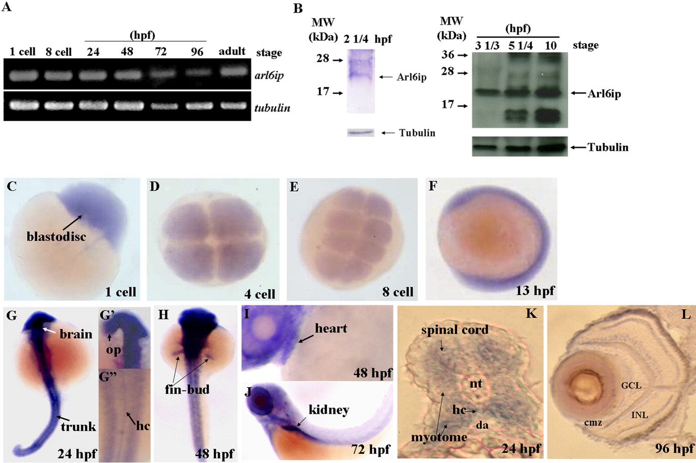
Fig. 1 Detection of the existence of arl6ip transcript and protein in zebrafish embryos using RT-PCR (A) and Western blot (B), respectively. The spatial expression of arl6ip mRNA was detected by whole-mount in situ hybridization (C-L). The arl6ip mRNA was first detected in blastodisc at the 1-cell stage (A,C), suggesting that the arl6ip transcript is maternally inherited. RT-PCR revealed that the arl6ip transcript was also detected at the 8-cell stage, 24, 48, 72, 96 hpf, and adulthood, when tubulin mRNA was used as a positive control (A). Total proteins were extracted from the wild-type embryos at 22 1/2, 3 1/3, 5 1/4 or 10 hpf, and the rabbit polyclonal antibody against ARMER was used to perform Western blot analysis when endogenous β-Tubulin was used as a control (B). Arl6ip was present at 2 1/4, 3 1/3, 5 1/4, and 10 hpf (indicated by arrow). Whole-mount in situ hybridization showed that the arl6ip was expressed ubiquitously in developing embryos at the 1-, 4-, and 8-cell stages and 13 hpf (C-F). The arl6ip was displayed in the brain, optic primordia (op), spinal cord, myotome, hypochord (hc), trunk, fin-bud, heart, kidney, and retina from 24 to 96 hpf (G-L). Dorsal view of arl6ip staining (G-G″,H). Lateral view of arl6ip expressions (F,I,J). arl6ip was expressed in retina by cross-sectioning of the eyes at 96 hpf (L). nt: notochord; da: dorsal aorta; GCL: ganglion cell layer; INL: inner nuclear layer; cmz: ciliary marginal zone; L: lens; hpf: hours post-fertilization of zebrafish embryos.
Image
Figure Caption
Figure Data
Acknowledgments
This image is the copyrighted work of the attributed author or publisher, and
ZFIN has permission only to display this image to its users.
Additional permissions should be obtained from the applicable author or publisher of the image.
Full text @ Dev. Dyn.

