
|
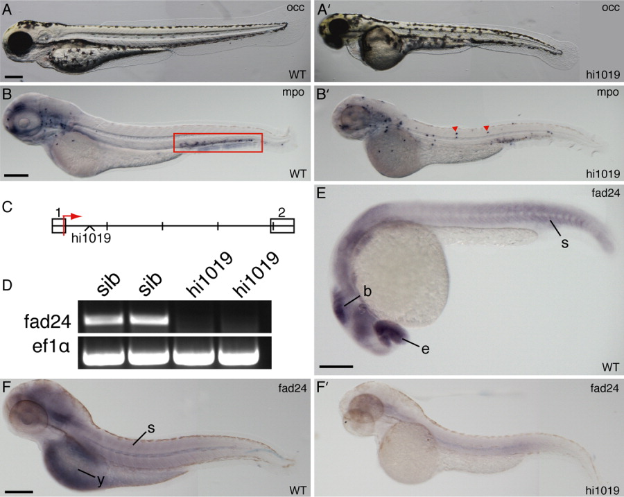
Fig. 1 Fad24hi1019 mutants exhibit a chronic inflammatory phenotype and reduced expression of fad24. A,A′: Oblique coherent contrast (OCC) images of WT and homozygous fad24hi1019 larvae at 3 dpf. B,B′: Zebrafish mpo in situ hybridizations of WT and fad24hi1019 larvae at 3 dpf. Box, CHT; arrows, neutrophils in the body of fad24hi1019 mutants. C: Genomic map of the first two exons of zebrafish fad24 showing the location of the hi1019 insertion; each section = 500 bp. D: RT-PCR analysis of WT (lanes 1,2) and fad24hi1019 mutant (lanes 3,4) single larvae at 3 dpf; ef1α is used as a control. E, F, F′: In situ hybridization of zebrafish fad24 in WT (E,F) and fad24hi1019 mutants (F′) at 26 hpf (E) and 3 dpf (F,F′). b, brain; e, eye; s, somite; y, yolk. Lateral view, anterior to the left. Scale bars = 200 μm. Representative results from at least three experiments with greater than 10 larvae per condition per experiment.