
Fig. 7
- ID
- ZDB-IMAGE-090106-18
- Genes
- Publication
- Jiang et al., 2008 - Exdpf is a key regulator of exocrine pancreas development controlled by retinoic acid and ptf1a in zebrafish
- All Figures
- Figures for Jiang et al., 2008
|
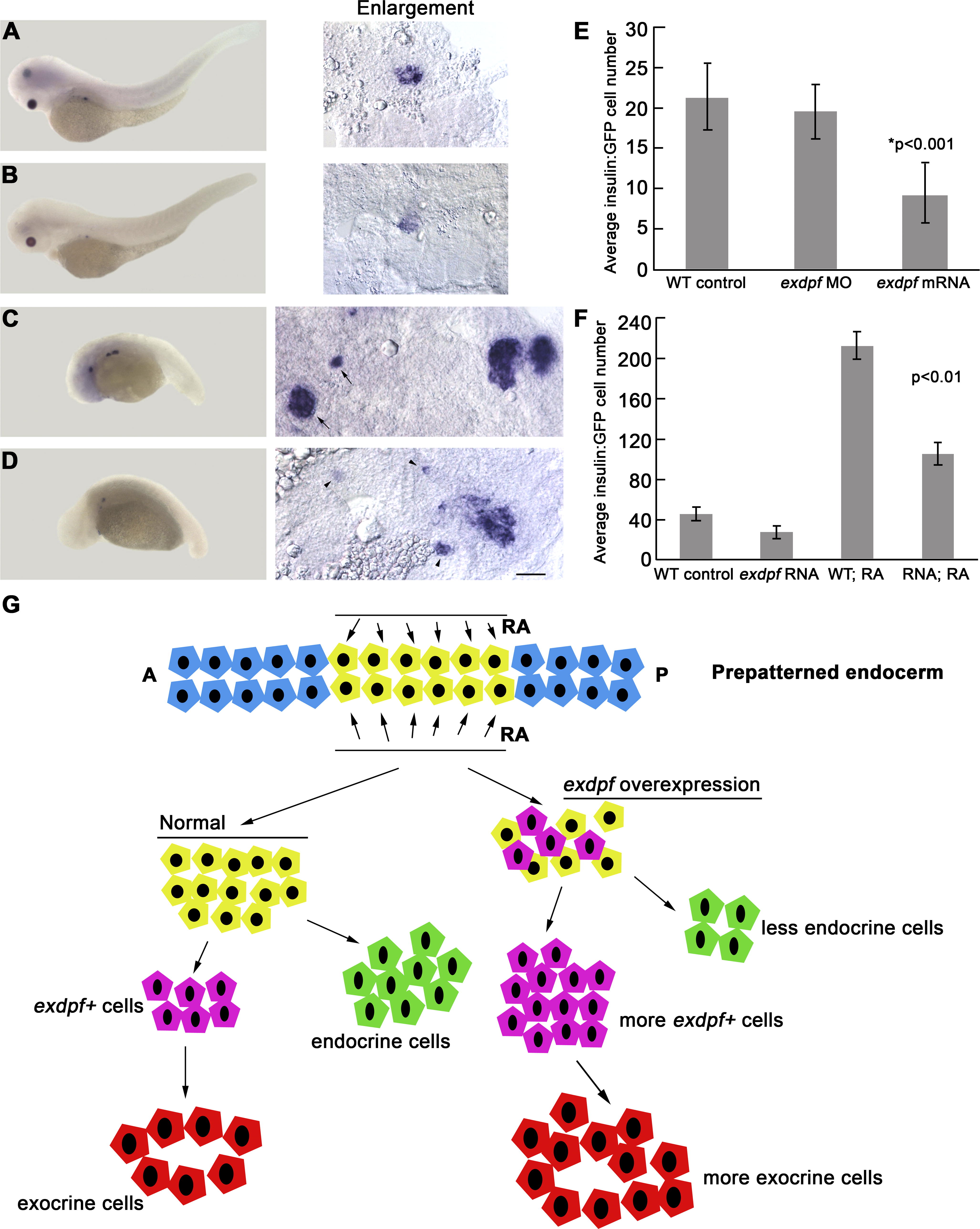
Fig. 7 Overexpression of exdpf Represses Endocrine Cell Differentiation in Both WT and RA-Treated embryos
(A–D) In situ hybridization on 3 dpf embryos using a preproinsulin probe. Enlargement: higher magnification of pancreatic area. (A) A WT embryo treated with DMSO as control. (B) A WT embryo injected with 100 pg of exdpf mRNA. (C) A WT embryo treated with 10-6 M of RA. Note anterior expansion of endocrine cells at the endogenous position and anterior ectopic endocrine cells (arrows). (D) A WT embryo injected with 100 pg of exdpf mRNA followed by RA treatment. Note weak anterior ectopic endocrine cells (arrowheads). Scale bar: 50 μm.
(E) A quantitative graph showing average GFP-positive cells per preproinsulin:GFP embryo at 24 hpf. Y axis, Mean ± SD.
(F) A quantitative graph showing average GFP-positive cells per preproinsulin:GFP embryo at 3 dpf. Y axis, Mean ± SD.
(G) A model for the effect of exdpf on endocrine cell differentiation. Normally, exdpf is only expressed in the exocrine cell precursors after endocrine cell differentiation. Injection of exdpf mRNA caused ectopic expression of exdpf in the committed pancreatic progenitors before endocrine cell differentiation. Therefore, more progenitors are transformed into exocrine cell fate and fewer progenitors develop into endocrine cells.