
|
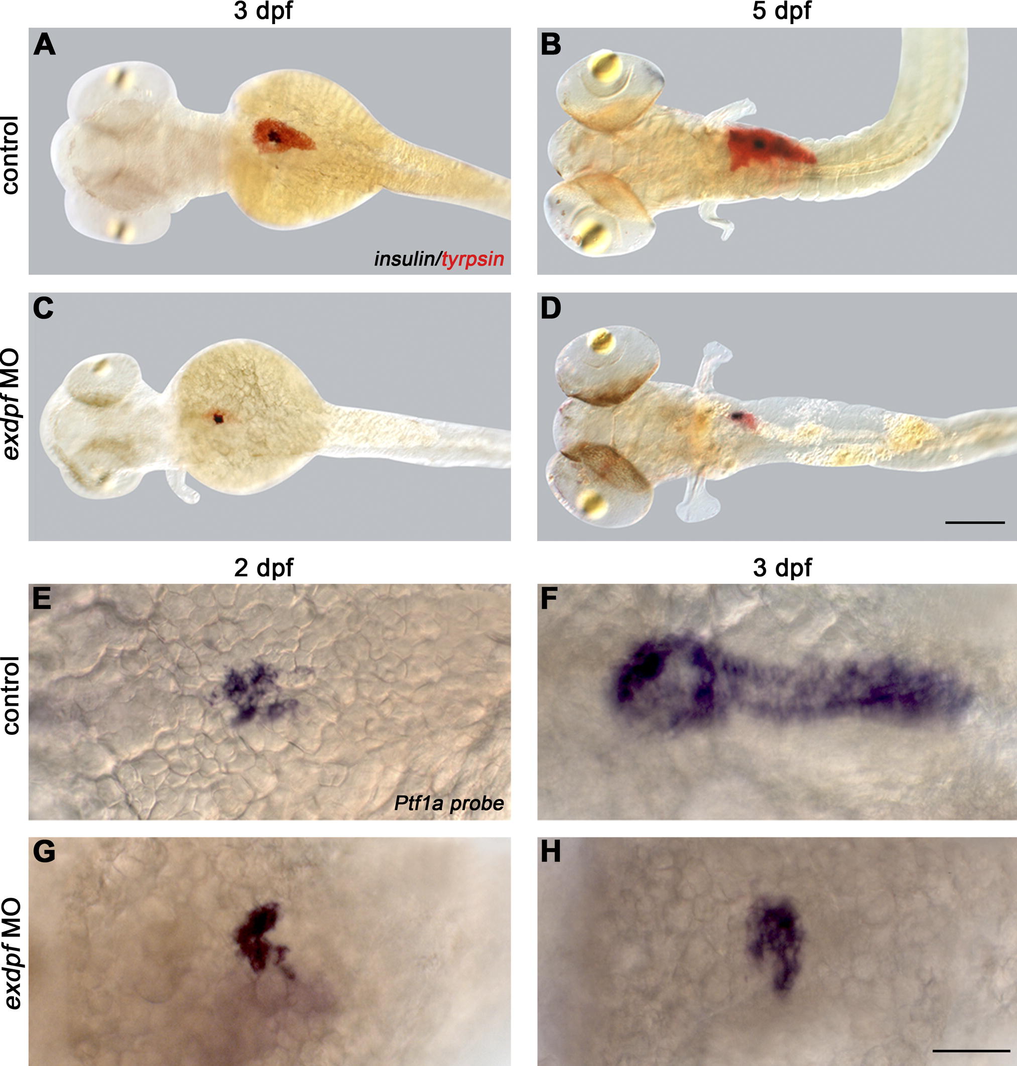
Fig. 2 Exdpf Is Required for the Exocrine Pancreas Differentiation and Expansion
(A–D) Double in situ hybridization using a trypsin probe (red) and a preproinsulin probe (blue). (A and B) Wild type (WT) embryos. (C and D) Embryos injected with exdpf morpholino. (A) A 3 dpf embryo. Note exocrine pancreas already expanded. (B) A 5 dpf embryo. Note both endocrine and exocrine mass increased. (C) An example of exdpf morphants, 3 dpf. Note exocrine mass is restricted to the anterior area, whereas endocrine portion appears normal. (D) An example of exdpf morphant at 5 dpf. Note exocrine pancreas is still restricted to the anterior area with limited increase in mass. All embryos are mounted in dorsal view, anterior to the left. Scale bar: 100 μm.
(E–H) In situ hybridization using a Ptf1a probe. Dorsal view, anterior to the left. (E and F) Wild type embryos at 2 dpf and 3 dpf, respectively. (G and H) Examples of exdpf morphants at 2 dpf and 3 dpf, respectively. Note Ptf1a expression is restricted to the anterior area at 2 dpf (G); the expression is still limited in the anterior area and the mass does not increase at 3 dpf (H). Scale bar: 50 μm.