
Image
|
Figure Caption
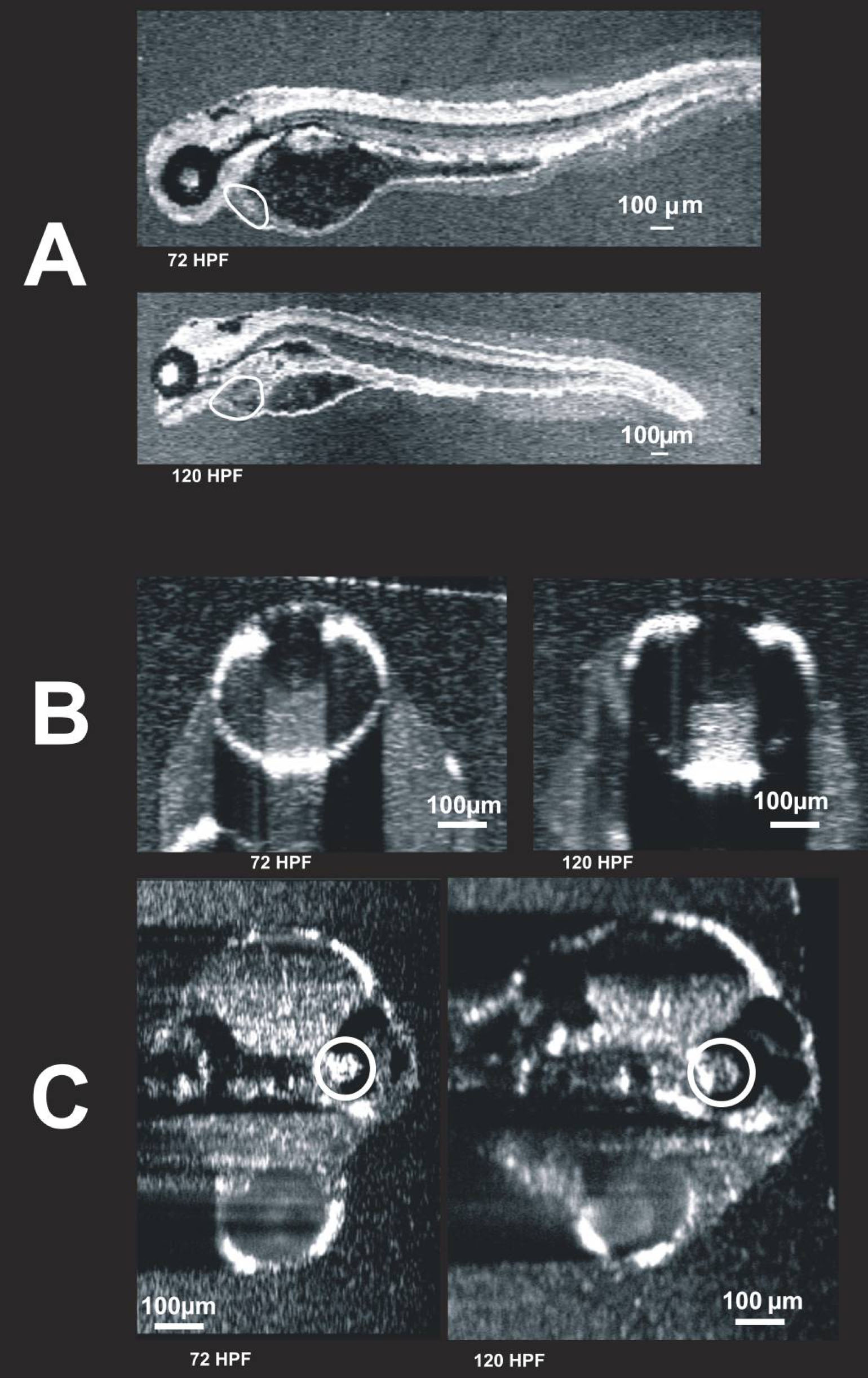
Fig. 10 Visualization of individual animals imaged on two different days. These images were obtained from the same embryo on two different days: 72 hpf and 120 hpf. C-mode images of the heart (A, circled), eye (B), and ear (C, circled) are presented. The heart is also visible in C, but blurred due to averaging over multiple cardiac cycles. It is possible that the first imaging session altered development. To compare the 120 hpf twice-imaged embryos to 120 hpf embryos imaged only once, refer to Figure 6 and Figure 9.
Acknowledgments
This image is the copyrighted work of the attributed author or publisher, and
ZFIN has permission only to display this image to its users.
Additional permissions should be obtained from the applicable author or publisher of the image.