
Image
|
Figure Caption
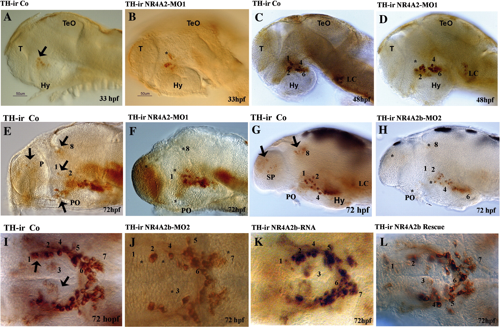
Fig. 4 TH expression during brain development in control and morphant larvae. Whole-mount views of TH protein expression (immunocytochemistry, brown staining), for all pictures anterior is to the left, in A–H dorsal is to the top I–L are ventral views. (A, B) Lateral view of 33 hpf embryos. (C, D) Lateral view of 48 hpf embryos. (E–H) Lateral view of 72 hpf larvae. (I–L) 72 hpf larvae viewed ventrally from the hypothalamus. (*) indicate missing THir expression in morphants (compare to black arrows in control panels).
Figure Data
Acknowledgments
This image is the copyrighted work of the attributed author or publisher, and
ZFIN has permission only to display this image to its users.
Additional permissions should be obtained from the applicable author or publisher of the image.
Reprinted from Molecular and cellular neurosciences, 39(4), Blin, M., Norton, W., Bally-Cuif, L., and Vernier, P., NR4A2 controls the differentiation of selective dopaminergic nuclei in the zebrafish brain, 592-604, Copyright (2008) with permission from Elsevier. Full text @ Mol. Cell Neurosci.