
|
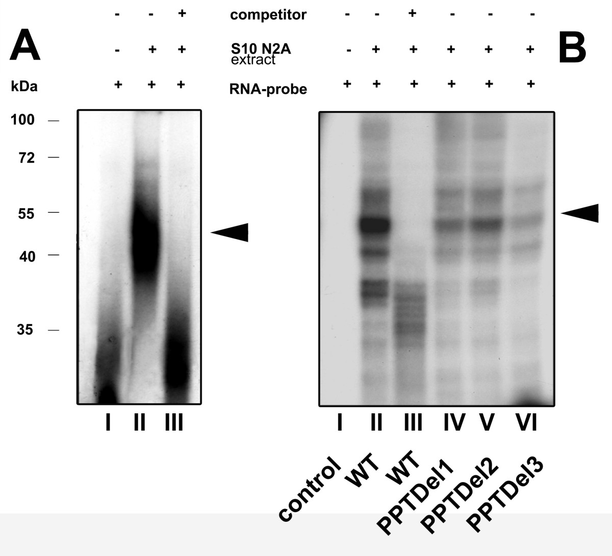
Fig. 5 RNA-EMSA reveals a RNA-protein complex on the IRES element: A) RNA-EMSA of wild type IRES. Internally labeled 32P RNA-probes were incubated with S10 extract from N2A cells. RNA-protein complexes, resolved on a 4% non-denaturing polyacrylamide gel were visualized by autoradiography. The presence of competitor (50 fold molar excess), S10 N2A extract and radiolabeled RNA probe is indicated by (+/-). The position of the RNA-protein binding complex is indicated by the arrow. B) UV cross-linking of RNA probes with S10 N2A extract: RNA-protein complexes were formed as indicated in (A) and samples were subjected to UV cross-linking followed by subsequent RNase treatment and resolved by 10% SDS PAGE. The composition of each sample is indicated as shown in A). Triangles on the right represent specific RNA-protein complexes and the numbers on the left represent a protein molecular weight marker kDa. Densitometrical analysis revealed a reduction to 39% (lane IV), 47% (lane V) and 27% (lane VI) with the wild type condition (lane II) set to 100%.