
Fig. 3
|
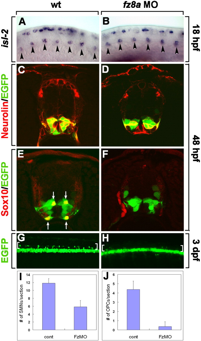
Fig. 3 fz8a function is required for motor neuron and oligodendrocyte development. A, B: Lateral views of spinal cords hybridized with an isl-2 RNA probe at 18 hpf, dorsal to the top and anterior to the left. A: Control embryos. B: fz8a MO-injected embryos. Arrowheads indicate primary motor neurons in the ventral spinal cord. C-F: Transverse section of the spinal cord of Tg(olig2:egfp) embryos, dorsal up. C, D: Combined anti-Neurolin (red fluorescence) and olig2:EGFP labeling in control embryos (C), and fz8a MO-injected embryos (D). E, F: Labeling with an anti-Sox10 antibody to mark OPCs (red fluorescence) in control (E) and fz8a MO-injected (F) Tg(olig2:egfp) embryos. G, H: Side views of whole-mount 3-dpf embryos, dorsal up and anterior to the left. G: Control Tg(olig2:egfp) embryo showing numerous olig2:EGFP+ OPCs in the dorsal spinal cord (brackets). H: fz8a MO-injected Tg(olig2:egfp) embryo. Only a few OPCs are evident in the dorsal spinal cord (brackets). I, J: Quantification of Neurolin+ secondary motor neurons (SMNs) (I) and Sox10+ OPCs (J) at 48 hpf in control (cont) and fz8a MO-injected (FzMO) embryos. Bars indicate the average number of cells per transverse section. Data were obtained from 10 sections from each of 5 control and 5 fz8a MO-injected embryos.