
|
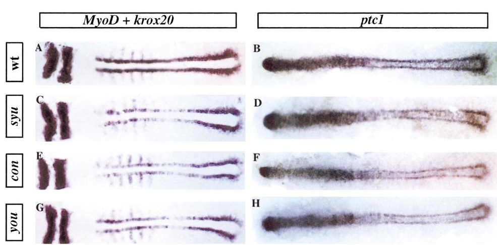
Fig. 2 Expression of ptc1 and MyoD in syu, con, and you at five to six somites. Dorsal flat preparations of embryos hybridised with probes for MyoD and krox-20 (A, C, E, and G) or ptc1 (B, D, F, and H). The krox 20 expression serves as a control for staining levels. (A and B) Wild-type embryos: note the strong expression of myoD in the adaxial cells and much weaker expression in the rostral somites. Expression of ptc1 overlaps myoD in the adaxial cells; note also the strong expression in the rostral neural tube. (C and D) syu, (E and F) con, (G and H) you; in all three mutants there is a slight reduction in the levels of expression of ptc1 and reduced and discontinuous expression of MyoD in the adaxial cells.
Reprinted from Developmental Biology, 216(2), Lewis, K.E., Currie, P.D., Roy, S., Schauerte, H., Haffter, P., and Ingham, P.W., Control of muscle cell-type specification in the zebrafish embryo by hedgehog signalling, 469-480, Copyright (1999) with permission from Elsevier. Full text @ Dev. Biol.