
Image
|
Figure Caption
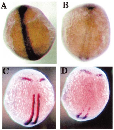
Fig. 1 Early expression of ptc1 and MyoD in yot homozygotes. yot homozygotes have reduced expression of ptc1 (B) and myoD (D) compared to wild type (A and C, respectively). ptc1 is still expressed throughout the adaxial cells (B) but at much lower levels than in wild-type (A), whereas MyoD is expressed only in the tailbud and most posterior adaxial cells (D). Note that the embryos were also hybridised with a probe for krox-20. The anterior stripe of expression that this reveals provides a spatial reference point and a control for staining levels.
Figure Data
Acknowledgments
This image is the copyrighted work of the attributed author or publisher, and
ZFIN has permission only to display this image to its users.
Additional permissions should be obtained from the applicable author or publisher of the image.
Reprinted from Developmental Biology, 216(2), Lewis, K.E., Currie, P.D., Roy, S., Schauerte, H., Haffter, P., and Ingham, P.W., Control of muscle cell-type specification in the zebrafish embryo by hedgehog signalling, 469-480, Copyright (1999) with permission from Elsevier. Full text @ Dev. Biol.