
Fig. 3
- ID
- ZDB-IMAGE-081104-12
- Genes
- Publication
- Jarinova et al., 2008 - Functional resolution of duplicated hoxb5 genes in teleosts
- All Figures
- Figures for Jarinova et al., 2008
|
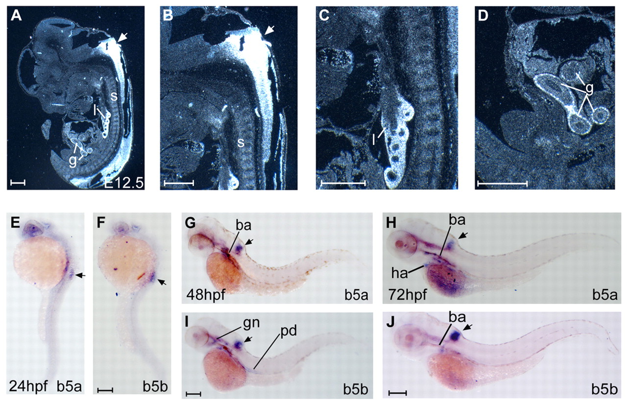
Fig. 3 Embryonic expression of mouse and zebrafish Hoxb5 genes. (A-D) Sagittal sections of E12.5 mouse embryos. The antisense Hoxb5 probe consisted of a 430 bp fragment from the Hoxb5 cDNA that includes the N-terminal part of the Hoxb5 protein (Krumlauf et al., 1987). (E-J) Whole-mount in situ hybridization on (E,F) 24 hpf, (G,I) 48 hpf and (H,J) 72 hpf zebrafish embryos using antisense riboprobes for hoxb5a (b5a, 550 bp probe) and hoxb5b (b5b, 600 bp probe). Zebrafish embryos are shown as lateral views. Arrows point to the embryonic neural tube. ba, branchial arches; g, gut; gn, trigeminal ganglion; l, lung; pd, pronephric duct; s, somites. Scale bars: 500 μm in A-D; 250 μm in E-J.