
|
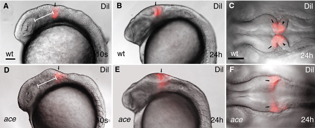
Fig. 6 Dil lineage-tracing reveals fate alteration of MHB primordial cell in ace mutants. All views are rostral to the left. (A,B,D,E) Lateral views; (C,F) dorsal views. (A-F) Labeling (red) wild-type and ace mutant embryos at equivalent rostrocaudal positions along the neuraxis reveals that the labeled cells in the mutants are not retained in the MHB compartment. The labeled mutant cells always end up at the caudal enlargement of the tectum (E,F). Arrows (A,B,D,E) point to the Dil-labeled group of cells. Arrows (C,F) point to the mesencephalic side of the labeled compartment; arrowheads (C) point to the hindbrain side of the Dil-labeled cell population. The white bar (A,F) shows the distance between the caudal edge of the otic vesicle and the Dil injection.