
Image
|
Figure Caption
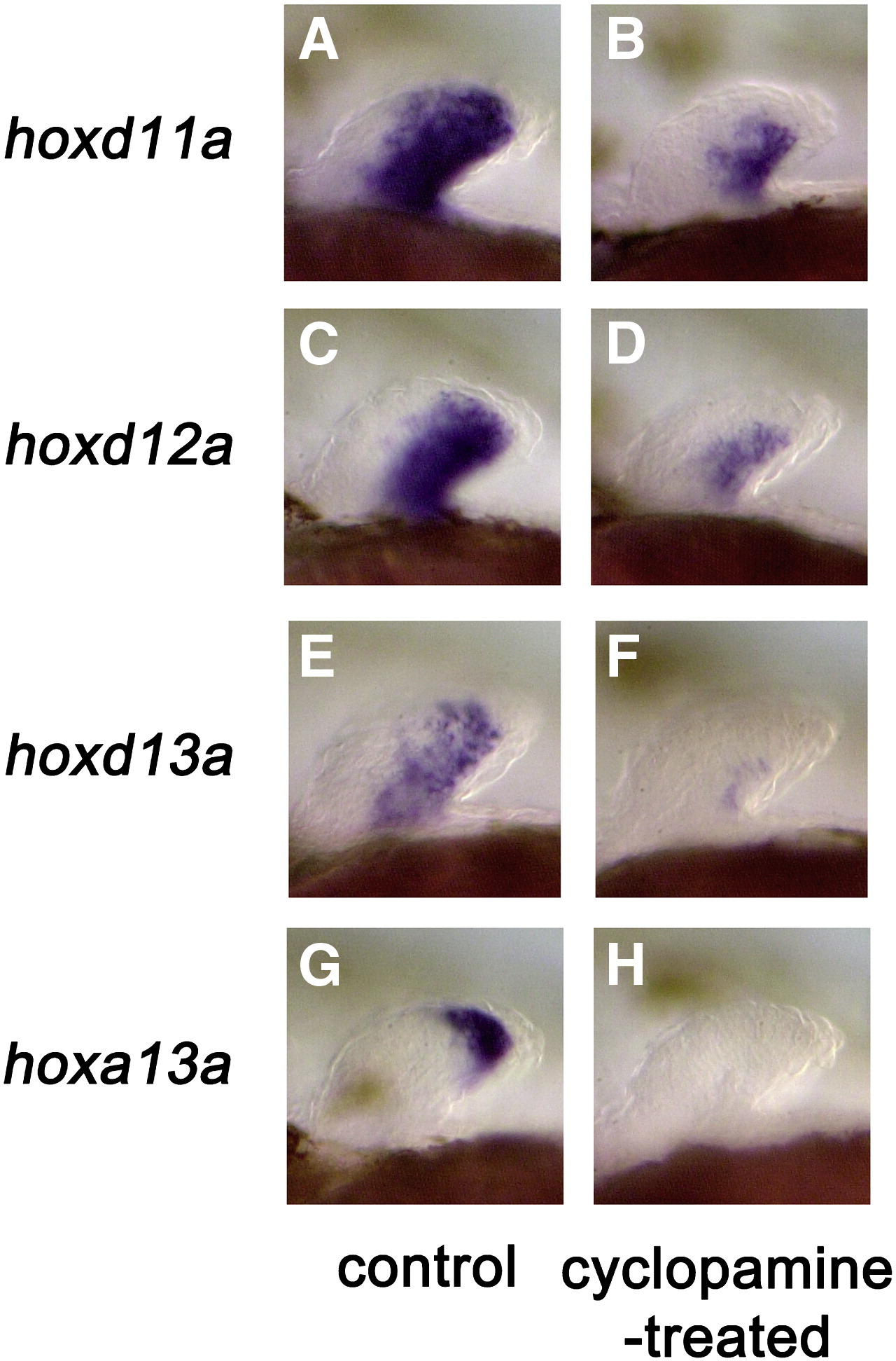
Fig. 6 Effect of cyclopamine treatment on hox gene expression in the pectoral fin bud. (A, C, E, G) Control embryos showing normal expression of hox genes. (B, D, F, H) Embryos treated with 50 μM cyclopamine for 12 h from 36 hpf. Note the complete absence of hox gene expression within the distal mesenchyme cells in cyclopamine-treated embryos. Lateral views of the left pectoral fins with anterior to the left in all panels. All embryos are approximately at 48 hpf. hpf: hours post fertilization.
Figure Data
Acknowledgments
This image is the copyrighted work of the attributed author or publisher, and
ZFIN has permission only to display this image to its users.
Additional permissions should be obtained from the applicable author or publisher of the image.
Reprinted from Developmental Biology, 322(1), Ahn, D., and Ho, R.K., Tri-phasic expression of posterior Hox genes during development of pectoral fins in zebrafish: Implications for the evolution of vertebrate paired appendages, 220-233, Copyright (2008) with permission from Elsevier. Full text @ Dev. Biol.