
Image
|
Figure Caption
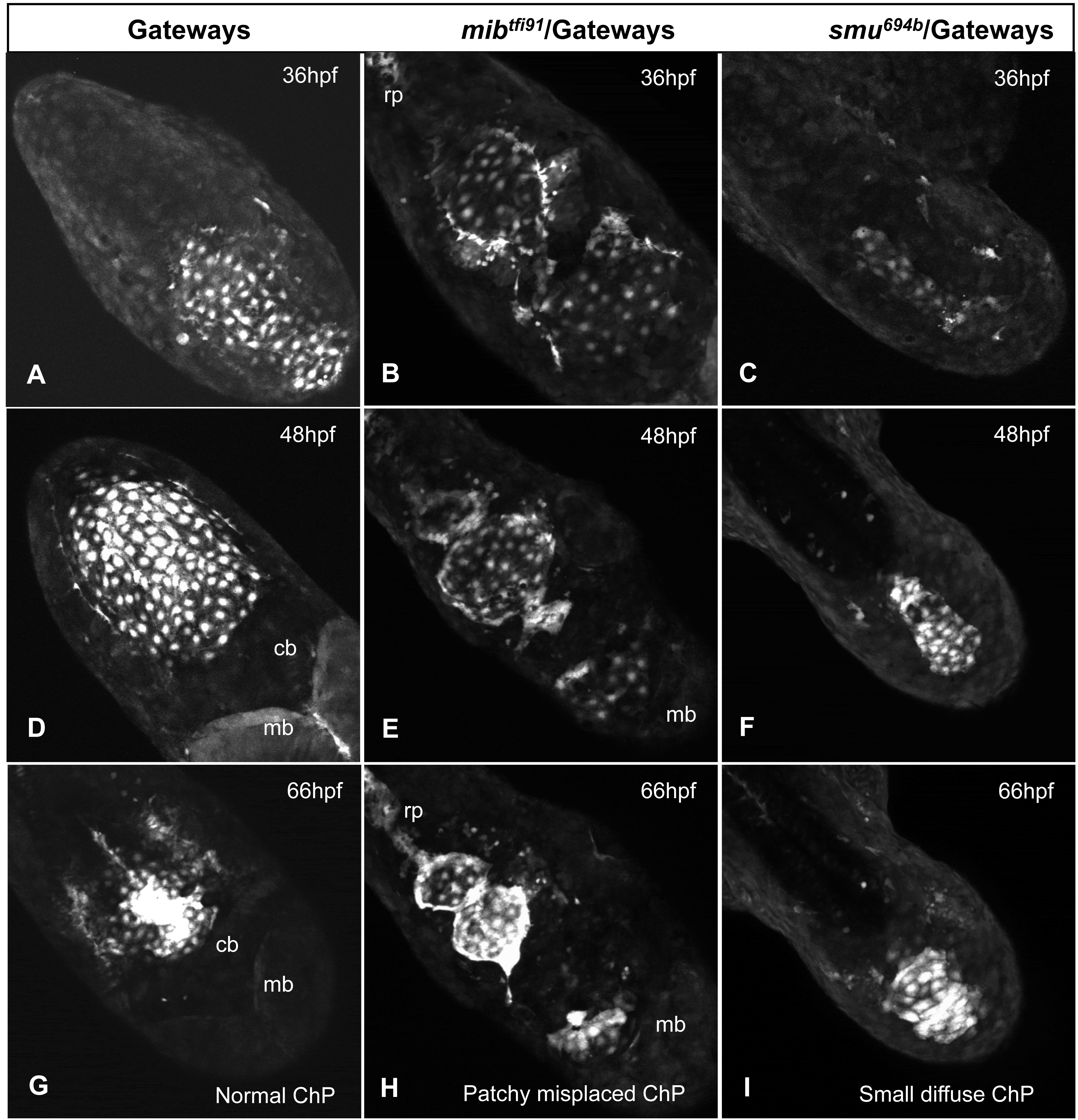
Fig. 5
Mutant analysis of the formation of the fourth ventricle ChP.
A, D, G – control; B, E, H, - mibtfi91; C, F, I - smu694b. A–C – 36hpf, D–F – 48hpf, G-I – 66hpf. Images are confocal z-projections extracted from the movies. All images are taken in dorsal view with the anterior part of the embryo towards the right-bottom corner. In controls and mib mutants dividing cells were detected and none of cells were fragmented. In smu a number of fragmenting cells were detected (purple arrow, movies) with no cells dividing. Abbreviations: cb – cerebellum, mb – midbrain, rp- roof plate.
Figure Data
Acknowledgments
This image is the copyrighted work of the attributed author or publisher, and
ZFIN has permission only to display this image to its users.
Additional permissions should be obtained from the applicable author or publisher of the image.
Full text @ PLoS One