
|
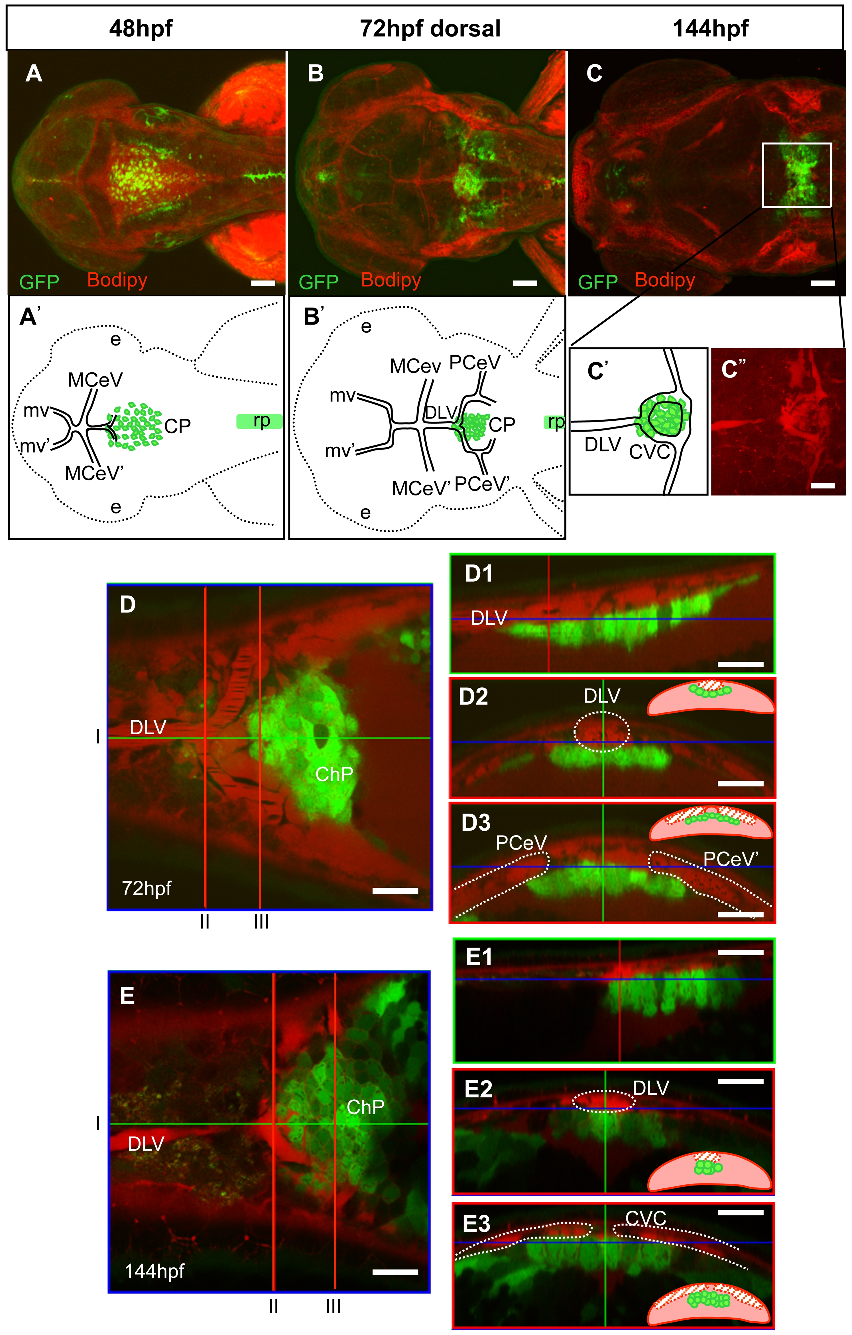
Fig. 4
Comparative analysis of formation of the fourth ventricle ChP and vasculogenesis.
Staining of Gateways by Bodipy (red) allows the in vivo analysis of ChP and cranial vasculature development. Extension of the DLV, growth of PCeV and closure of CCV occur at the same time as the transformation of the tela choroidea into ChP. A–C, confocal images at three different time points with their respective explicative drawings (A′, B′, C′, C″). D, E - dorsal optical section focused on ChP; D1 and E1 saggital optical sections at the level of the green line in D, E; D2, D3, E2, E3 cross optical sections at the level of the red lines in D, E. At all times ChP keeps a close contact with the developing vessels. Abbreviations: ChP – choroid plexus, CVC – choroidal vascular circuit, dlv- dorsal longitudinal vein, mv- midbrain vein, PCeV- posterior cerebral vein, rp- roof plate. Scale bar in D, D1–D3, E, E1–E3, 20 μm.