
Image
|
Figure Caption
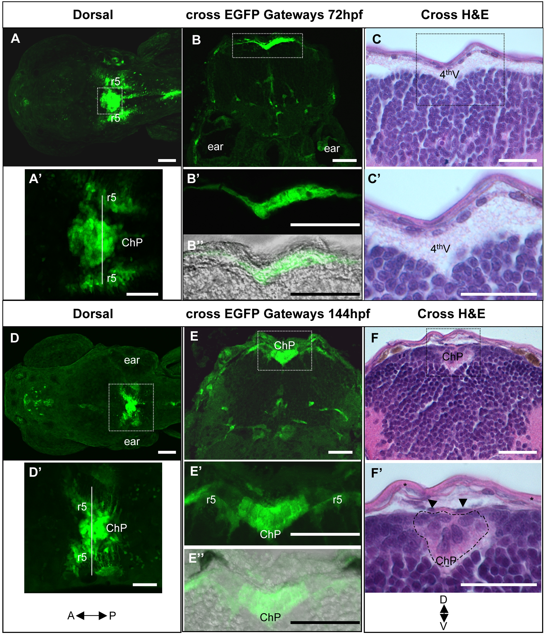
Fig. 2
Comparative histological and developmental analysis of the fourth ventricle choroid plexus in Gateways zebrafish transgenics.
A–C – 72hpf, D–F – 144hpf. A, A′, D, D′, - dorsal view, B, B′, B″, C, C′, E, E′, E″, F, F′ – cross sections. All A, B, D, E images show GFP expression, all C and F bright field images show staining with hematoxylin-eosin; B″ and E″ are composite fluorescence-bright-field images. Abbreviations: ChP – choroid plexus, 4thV – fourth ventricle, r5 – rhombomere 5, starlet in F′ – skin epithelium, arrowheads in F′ – vessel.
Figure Data
Acknowledgments
This image is the copyrighted work of the attributed author or publisher, and
ZFIN has permission only to display this image to its users.
Additional permissions should be obtained from the applicable author or publisher of the image.
Full text @ PLoS One