
Image
|
Figure Caption
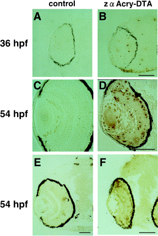
Fig. 8 Apoptosis of DTA-expressing embryos. In situ TUNEL analysis was done by using central, transverse frozen sections of zebrafish embryonic eyes. DTA-expressing or control embryos at 36 and 54 hpf were embedded in OCT compound and frozen sectioned. FITC conjugate–dUTP-labeled cells were visualized with anti-FITC-horseradish peroxidase conjugate antibody and DAB. Samples were examined under Normarsky microscopy (Axioplan; Carl Zeiss). Scale bars, 50 μm.
Acknowledgments
This image is the copyrighted work of the attributed author or publisher, and
ZFIN has permission only to display this image to its users.
Additional permissions should be obtained from the applicable author or publisher of the image.
Reprinted from Developmental Biology, 255(1), Kurita, R., Sagara, H., Aoki, Y., Link, B.A., Arai, K.-I., and Watanabe, S., Suppression of lens growth by alphaA-crystallin promoter-driven expression of diphtheria toxin results in disruption of retinal cell organization in zebrafish, 113-127, Copyright (2003) with permission from Elsevier. Full text @ Dev. Biol.