
Image
|
Figure Caption
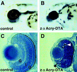
Fig. 3 Morphological characterization of the zα Acry-DTA-expressed embryo. Views of live wild-type (A) and DTA-expressing (B) embryos at 54 hpf. Toluidine blue-stained sections through control (C) and DTA-expressing (D) eyes at 54 hpf. The three nuclear cell layers, retinal ganglion layer (G), inner nuclear layer (I), and outer nuclear layer (O), are visible in the wild-type retina. No layers are apparent in DTA-expressing retina, and apoptotic cells are indicated by arrowheads (D). Scale bars, 20 μm.
Acknowledgments
This image is the copyrighted work of the attributed author or publisher, and
ZFIN has permission only to display this image to its users.
Additional permissions should be obtained from the applicable author or publisher of the image.
Reprinted from Developmental Biology, 255(1), Kurita, R., Sagara, H., Aoki, Y., Link, B.A., Arai, K.-I., and Watanabe, S., Suppression of lens growth by alphaA-crystallin promoter-driven expression of diphtheria toxin results in disruption of retinal cell organization in zebrafish, 113-127, Copyright (2003) with permission from Elsevier. Full text @ Dev. Biol.