
|
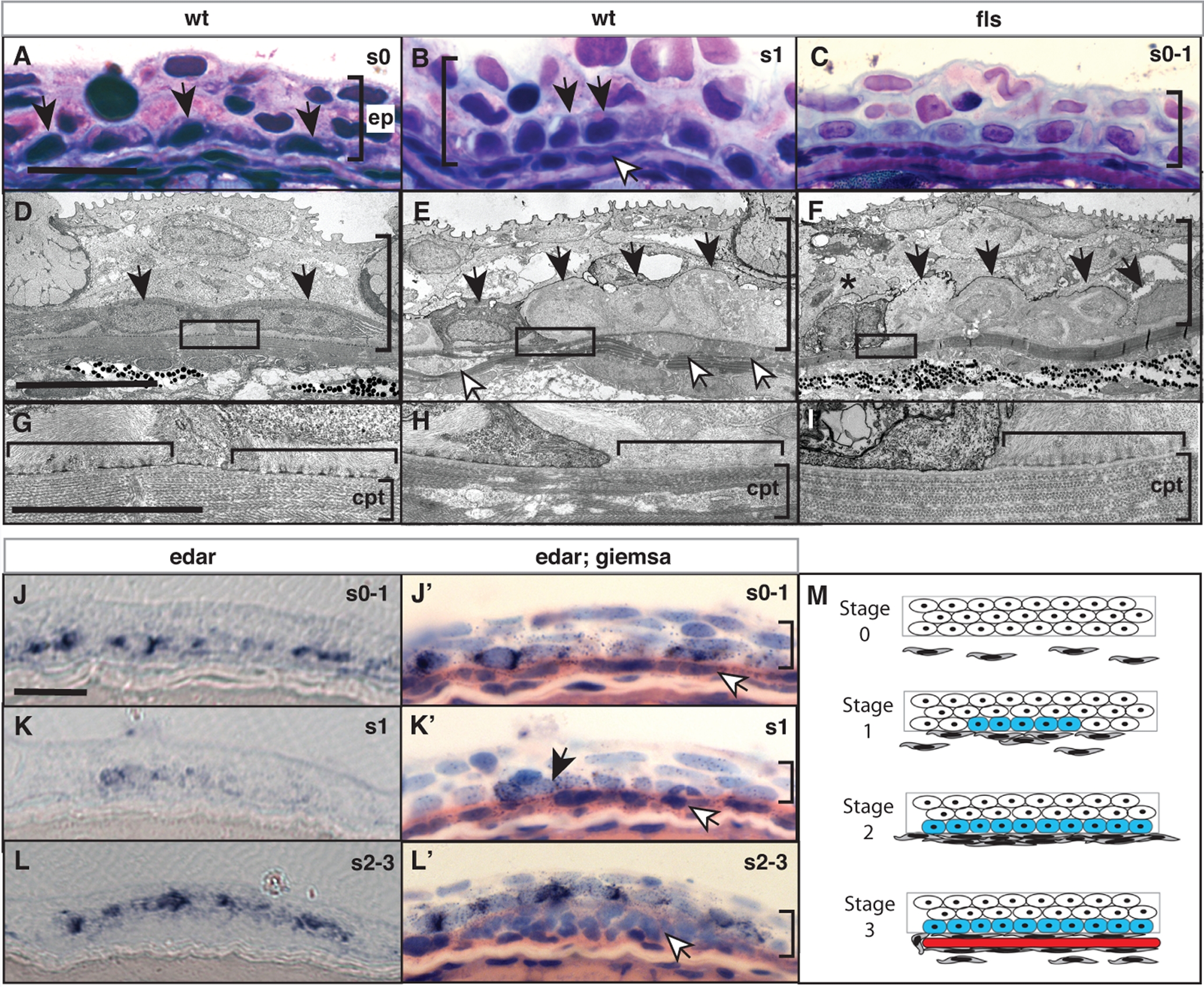
Fig. 5 Eda signaling regulates the formation of an epidermal placode during scale development.
Histological analysis of wild type (A, D, B, E) and flste370f (C, F, I) integument of 8 mm standard length. In wild type juveniles (B, E), basal epidermal cells (black arrow heads) show a heightened, and cuboidal morphology at sites of scale development as indicated by an accumulation of migrating fibroblast-like cells (white arrowheads). (H) This morphology of the epidermis is associated with a reworking of the collagen layer of the stratum compactum (cpt; [24]). This is in contrast to the flattened morphology of basal epidermal cells lateral to those of the scale placode (A, D) and underlying dense stratum compactum (G). In flste370f this basal epidermal structure is disorganized and cell morphology is disrupted (C, F) including evidence of cell death (asterisk). The lack of reworking of the collagen of the stratum compactum in the flste370f mutant is associated with retention of hemidesmosomes (horizontal bracket G–I). edar is expressed in cells of the wildtype epidermis (J, K, L). Counterstaining of the same sections confirms the expression in basal cells overlying initial accumulating fibroblasts (white arrowheads; J′, K′, L′). Expression of edar is observed prior to organization of the placode and fibroblast aggregation and maintained in cells of the epidermal placode through early scale development (J–L). M) Schematic depicting scale development and edar expression. The stages of scale development are modeled using analogous stages as described for hair development [35]; stage 0, nascent epidermis; stage 1, placode specification; stage 2, scale pocket; stage 3, matrix deposition and ossification. Blue, edar expression; red, scale formation. ep, epidermis; cpt stratum compactum. The vertical bracket demarcates the extent of the epidermis in the sections. Measurement bar equals 10 μm.