
|
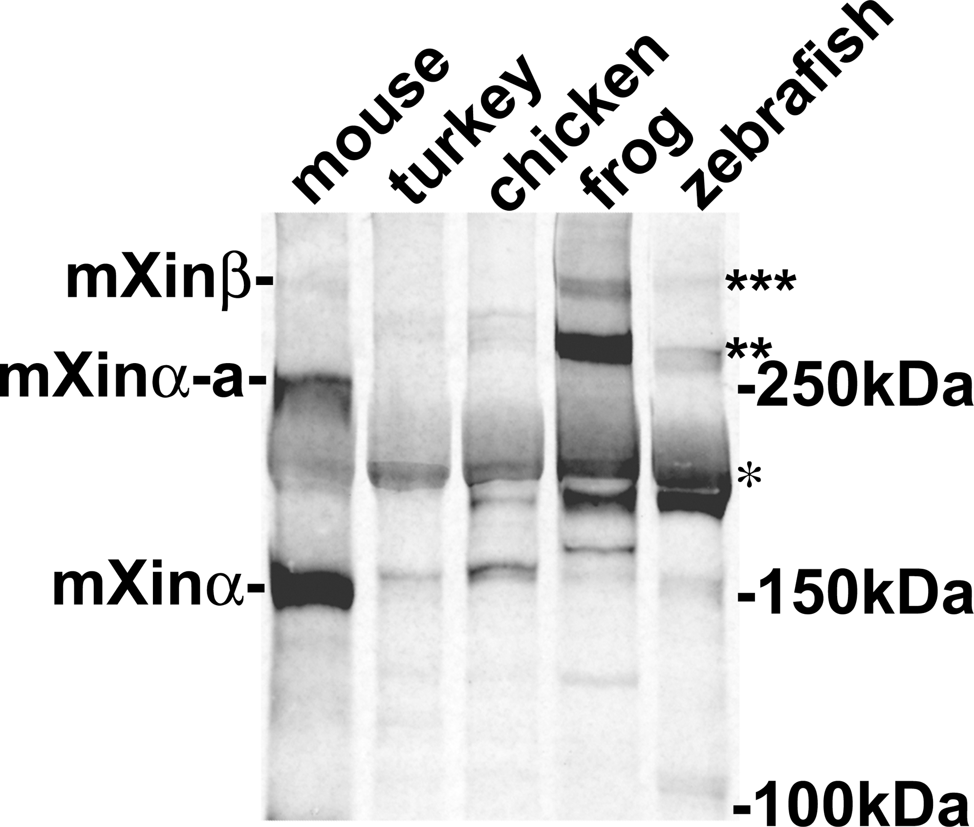
Fig. 4
Western blot analysis of protein extracts prepared from mouse, turkey, chicken, frog and zebrafish hearts with polyclonal U1013 anti-Xin antibody.
As previously reported [5], the U1013 antibody generated against the N-terminal fragment of mXinα including the Xin repeat region specifically reacts with mXinα (155 kDa), mXinα-a (250 kDa) and mXinβ (∼340 kDa) from mouse heart. Similarly, this antibody recognizes a 217 kDa band (indicated by *), and 280–295 kDa bands (indicated by**) from turkey, chicken, frog and zebrafish heart extracts. These bands may represent Xinα and its splicing variants Xinα-a. Many degraded fragments were also detected by this antibody. In frog and zebrafish but not turkey and chicken heart extracts, this antibody also reacts with a 335 kDa band (indicated by ***), which may represent Xinβ isoform.