
Image
|
Figure Caption
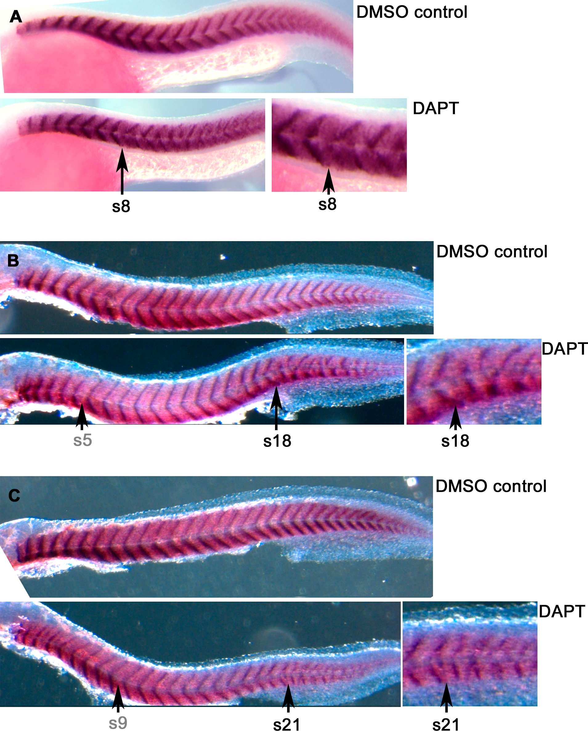
Fig. 2
Blocking Notch Signalling Causes Somite Boundary Defects after a Long Delay
Embryos were treated with 100 μm DAPT or with DMSO (control) medium and stained by ISH for titin at the end of somitogenesis to reveal somite boundaries. Treatment was begun (A) at 3 hpf, (B) at 5-somite stage, or (C) at 9-somite stage. Arrows with grey labels indicate stage at onset of DAPT treament; arrows with black labels indicate the level of the earliest defective somite. A detailed view of the region where disruption begins is shown to the right of each DAPT specimen.
Figure Data
Acknowledgments
This image is the copyrighted work of the attributed author or publisher, and
ZFIN has permission only to display this image to its users.
Additional permissions should be obtained from the applicable author or publisher of the image.
Full text @ PLoS Genet.