
Image
|
Figure Caption
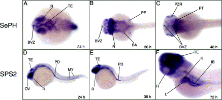
Fig. 7 Expression patterns of selenoproteins SePH (A–C) and selenophosphate synthetase SPS2 (D–F) in zebrafish embryos. The development stage is indicated in the right corner of each panel in hours post-fertilization. BVZ, brain ventricular zone; IB, intestinal bulb; K, kidney; L, liver; MY, myotomes; OV, olfactory vesicle; PD, pronephric duct; PF, pectoral fins; PT, posterior tectum; PZR, proliferative zone of the retina; R, retina; TE, tectum.
Acknowledgments
This image is the copyrighted work of the attributed author or publisher, and
ZFIN has permission only to display this image to its users.
Additional permissions should be obtained from the applicable author or publisher of the image.
Reprinted from Gene expression patterns : GEP, 3(4), Thisse, C., Degrave, A., Kryukov, G.V., Gladyshev, V.N., Obrecht-Pflumio, S., Krol, A., Thisse, B., and Lescure, A., Spatial and temporal expression patterns of selenoprotein genes during embryogenesis in zebrafish, 525-532, Copyright (2003) with permission from Elsevier. Full text @ Gene Expr. Patterns