
|
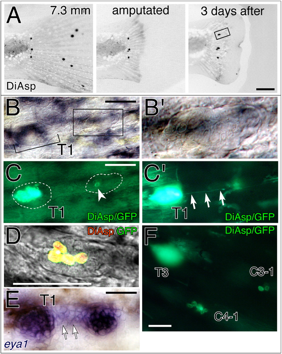
Fig. 5 Accessory neuromast formation of the terminal neuromasts in zebrafish. A: Embryos were analyzed 3 days after amputation of the caudal fin. B,B′: The boxed region of the lower panel in A is shown under differential interference contrast (DIC) optics. Higher magnification of the regenerated caudal lateral line system (CLL) neuromast is shown in B′. C,C′: The same region as B is shown under fluorescent illumination (C) with a deeper focal plane (C′). Positions of the founder neuromast and the budding neuromast are indicated by dotted lines. Arrowhead indicates DiAsp-positive hair cells. Arrows indicate the extending lateral line efferent nerve. D: Confocal image of a budding neuromast. The fluorescent image is superimposed over the DIC image. E: The same sample as A-C was analyzed by in situ hybridization using eya1 RNA probe. Eya1 mRNA is expressed in the cells between the founder neuromast and the regenerated neuromast (arrows). F: Example of a single terminal neuromast (T3) extending two prospective CLL neuromasts (C3-1 and C4-1) during the regeneration process. Scale bars = 500 μm in A, 50 μm in B-F.