
|
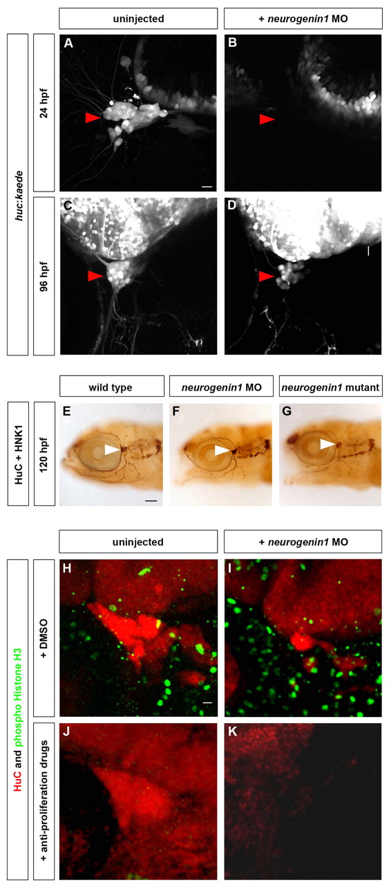
Fig. 6 The trigeminal sensory ganglia of neurogenin1 mutant and morphant embryos are solely formed from late-born neurons. Neurogenin1-depleted embryos develop smaller trigeminal sensory ganglia formed from late-born neurons only. (A-D) Embryos carrying the huc:kaede transgene were injected with 6 ng of neurogenin1 antisense morpholino (B,D) or uninjected (A,C). At 24 hpf, the trigeminal sensory ganglia are visible in uninjected embryos by the expression of Kaede (A,C) but no trigeminal sensory neurons are detectable in the neurogenin1 morphants at 24 hpf (B). At 96 hpf, the trigeminal sensory ganglia are visible in neurogenin1 morpholino-injected embryos (D) but contain fewer neurons than uninjected embryos (C). Side view, anterior towards the left. Scale bar: 10 μm. (E-G) The morphology of the neurons of the trigeminal sensory ganglia was analyzed by immunostaining in wild-type (E), neurogenin1 morphant (F) and neurogenin1 mutant (G) embryos with HuC, a pan-neuronal marker, and HNK-1, a marker labeling the cell surface of sensory neurons. White arrowheads indicate the trigeminal sensory ganglia. Side view, anterior towards the left. Scale bar: 100 μm. (H-K) To determine whether the trigeminal sensory ganglia in neurogenin1 morphant embryos are partly formed from early-born neurons, embryos were treated with 2% DMSO alone (H,J) or with 20 mM hydroxyurea and 150 μM aphidicolin (I,K) at 24 hpf. HuC staining (red) labels the trigeminal sensory ganglia. Staining for the mitotic marker phospho-histone H3 (green) was used to monitor the number of proliferating cells in the whole embryos. Proliferation was not affected in mock-treated embryos (H,J) but was significantly reduced in treated embryos (I,K). No trigeminal sensory neurons are detectable in the neurogenin1 morphant embryos treated with the anti-proliferative drugs (K), in contrast to the mock-treated neurogenin1 morphant (I) or wild-type embryos (H,J). Side view, anterior towards the left. Scale bar: 10 μm.