
|
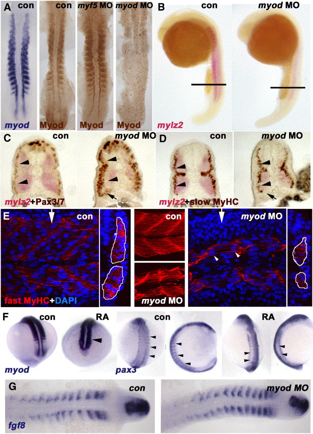
Fig. 6 Myod is required for normal fast muscle differentiation and suppression of Pax3/7. In situ mRNA hybridisation for myod, mylz2 myosin light chain, fgf8 or pax3 or immunodetection of Myod, Pax3/7, slow or fast MyHC viewed in dorsal flatmount (A, G 15 s), lateral wholemount (B, 26 s dorsal to right), transverse section of wholemount (C, D, 26 s dorsal to top), confocal stack (E, middle) or section in lateral view (E, 24 hpf medial somite anterior to left, dorsal at top, with transverse projection at white arrows shown to right) and wholemount (F, mid somitogenesis, anterior to top). (A) Myod-specific immunoreaction co-localises with myod mRNA. Injection of myod morpholino oligonucleotide (MO) ablates Myod protein, whereas myf5 MO enhances Myod immunoreaction. (B-D) Myod MO reduces mylz2 mRNA in fast muscle (B-D), without affecting slow muscle differentiation (D). Cryosections at the level indicated in panel B reveal reduced lateral migration of slow fibres (D), correlating with increased lateral Pax3/7-expressing cells (C). Pax3/7 expression in neural tube/crest appears unaffected. Note that all fast fibres appear medial to slow fibres, that most Pax3/7+ cells lie lateral to slow fibres (arrowheads C, D), but that Pax3/7+ cells accumulate ventrally next to the residual fast muscle (arrows C, D). (E) Similarly, myod MO reduces the quantity of muscle labelled for fast MyHC (outlined in white in transverse projections). Note that some fibres still appear multinucleate (arrowheads). (F) Exposure of embryos to 10- 7 M retinoic acid up-regulates myod mRNA in lateral presomitic mesoderm and somite (arrowhead, left pair, 15 s dorsal view of tailbud), RA reduces pax3 mRNA in nascent somites of 13 s embryos (arrowheads, shown at left in dorsal view and to right in lateral view). Note the up-regulation of pax3 signal in neural tissue. (G) Myod MO has no effect on fgf8 mRNA accumulation.
Reprinted from Developmental Biology, 302(2), Hammond, C.L., Hinits, Y., Osborn, D.P., Minchin, J.E., Tettamanti, G., and Hughes, S.M., Signals and myogenic regulatory factors restrict pax3 and pax7 expression to dermomyotome-like tissue in zebrafish, 504-521, Copyright (2007) with permission from Elsevier. Full text @ Dev. Biol.