
|
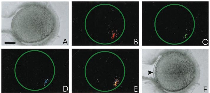
Fig. 8 A representative example of a zebrafish egg with fluorescent microspheres loaded into its periphery. A and F are bright-field images taken before and after activation, respectively. A shows no separation between the chorion and the egg plasma membrane, whereas in F, the chorion is clearly raised (see arrowhead). B to D indicate confocal images taken at an optical plane containing the fluorescent microspheres. The fluorescent image in B was taken before the addition of 0.5% fructose in egg-water and then C and D at 2.5 and 5 min later, respectively. E shows the fluorescent images from B, C, and D superimposed on one another. This demonstrates that the egg does not rotate within its expanding chorion during the activation process. Scale bar is 200 μm.
Reprinted from Developmental Biology, 214(1), Lee, K.W., Webb, S.E., and Miller, A.L., A wave of free cytosolic calcium traverses zebrafish eggs on activation, 168-180, Copyright (1999) with permission from Elsevier. Full text @ Dev. Biol.