
|
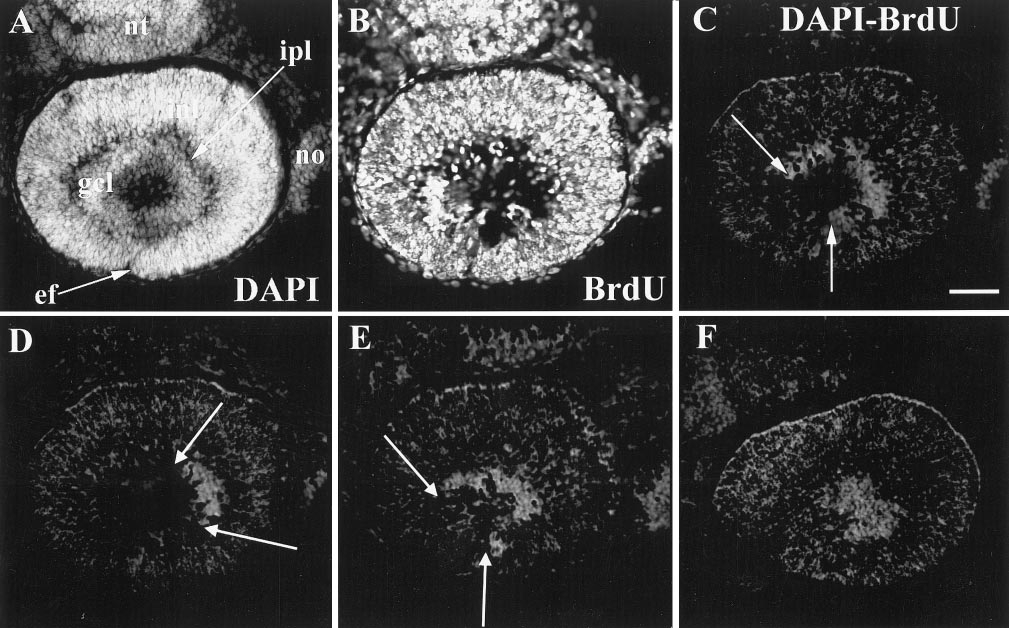
Fig. 3 The patch of ganglion cells advanced around the embryonic fissure in an arc, beginning in ventronasal retina. 31 hpf BrdU, 41 hpf sacrifice. A and B are the same section, stained as indicated. (C) Difference image of A and B, showing the arc of unlabeled cells in the ganglion cell layer, the two borders indicated by arrows. (D, E, and F) Similar difference images (DAPI and BrdU) from adjacent sections from this same retina to show that the unlabeled cells extend into lateral (D) and more medial (E, F) sections to form a disk of unlabeled cells. Abbreviations: as in Figs 1 and 2; nt, neural tube. Calibration: 40 μm.
Reprinted from Developmental Biology, 207, Hu, M. and Easter, Jr., S.S., Retinal neurogenesis: the formation of the initial central patch of postmitotic cells, 309-321, Copyright (1999) with permission from Elsevier. Full text @ Dev. Biol.