
|
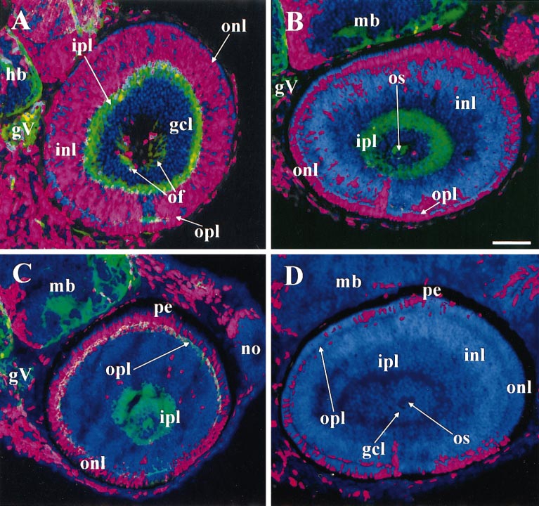
Fig. 7 The ventronasal retina was the most precocious region for both inner and outer nuclear layers as well as the ganglion cell layer. All panels labeled with DAPI (blue), BrdU (red), and (except for D) zn12 (green). (A) 38 hpf BrdU, 52 hpf sacrifice. A patch of BrdU-negative cells appears in ventronasal retina with abundant zn12 labeling in the adjacent outer plexiform layer, evidence for precocious birthdates and differentiation. (B) 43 hpf BrdU, 57 hpf sacrifice. Most of the inner nuclear layer is BrdU-negative. (C) 48 hpf BrdU, 62 hpf sacrifice. A patch of BrdU-negative cells has now appeared in ventronasal retina adjacent to the most heavily zn12-positive patch of the outer plexiform layer, evidence for precocious birthdates and differentiation. (D) 54 hpf BrdU, 68 hpf sacrifice. No zn12 staining. The outer nuclear layer contains BrdU-negative cells everywhere, but small patches of BrdU-positive cells remain, disconnected with one another. For this reason, a summary drawing, such as the one in Fig. 6, cannot be made for the INL and ONL. Abbreviations: as in Figs. 1–6, hb, hindbrain; mb, midbrain; pe, pigmented epithelium. Calibration: 40 μm.
Reprinted from Developmental Biology, 207, Hu, M. and Easter, Jr., S.S., Retinal neurogenesis: the formation of the initial central patch of postmitotic cells, 309-321, Copyright (1999) with permission from Elsevier. Full text @ Dev. Biol.