
Image
|
Figure Caption
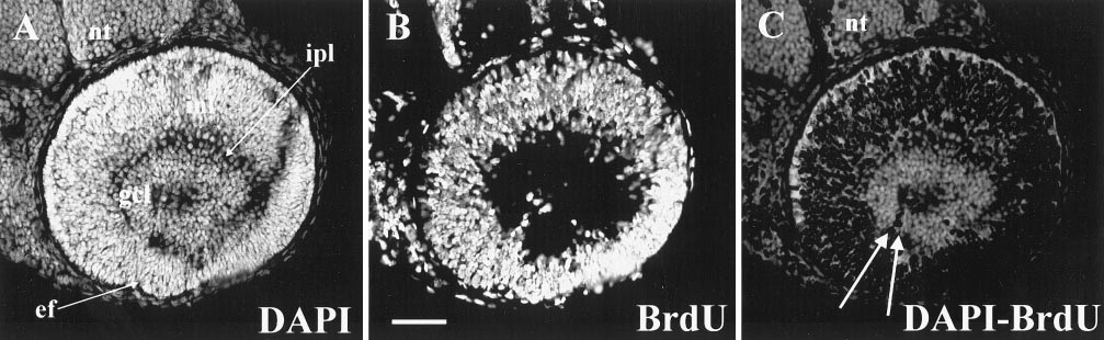
Fig. 4 A circle of ganglion cells. 37 hpf BrdU, 51 hpf sacrifice. A and B stained as indicated. (C) Difference image shows a nearly complete circle of BrdU-negative cells with the ventronasal and ventrotemporal edges separated by a thin row of proliferative (BrdU-positive) cells that mark the embryonic fissure. Abbreviations: as in Figs 1–3. Calibration: 40 μm.
Acknowledgments
This image is the copyrighted work of the attributed author or publisher, and
ZFIN has permission only to display this image to its users.
Additional permissions should be obtained from the applicable author or publisher of the image.
Reprinted from Developmental Biology, 207, Hu, M. and Easter, Jr., S.S., Retinal neurogenesis: the formation of the initial central patch of postmitotic cells, 309-321, Copyright (1999) with permission from Elsevier. Full text @ Dev. Biol.