
|
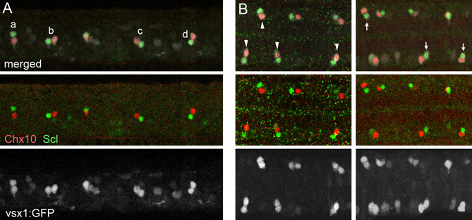
Fig. S6 The final location of the V2a and V2b neurons is not fixed. Double immunohistochemical staining of Chx10 (red) and Scl (green) in Tgvsx1:GFP embryos (GFP signal is white) at 20 hpf. (A) Lateral view. (B) Dorsal view. V2a/V2b pairs show various configurations. For example, in the lateral view (A), the relative position of Scl-positive cells to their sibling Chx10-positive cells could be to the dorsal (a), to the anterior (b), to the posterior (c) and to the ventral (d). Similarly, in the dorsal view (B), the relative position of Scl-positive cells could be to the lateral (arrowheads) and to the medial (arrows).