
|
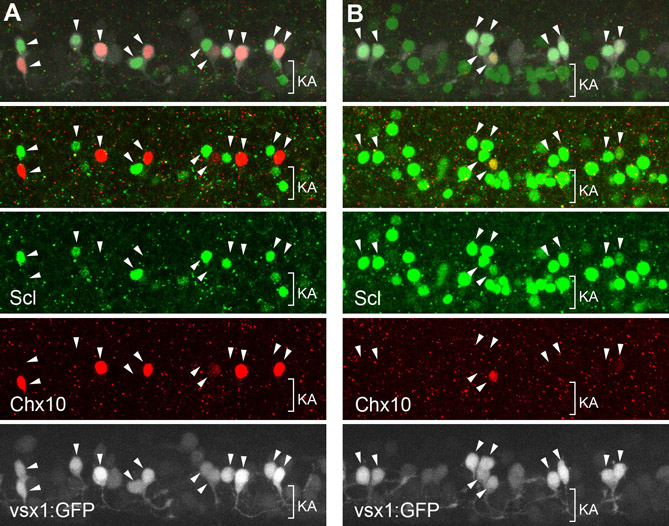
Fig. S3 Notch-signaling affects the cell fate of V2a andV2b neurons. Double immunohistochemical staining of Chx10 (red) and Scl (green) in Tgvsx1:GFP embryos (GFP signal is white) at 24 hpf. (A) Control experiment (Tgvsx1:GFP alone). (B) Notch1a intracellular fragment (NICD, an activated form of Notch1a) was widely expressed in neurons following a cross between TgUAS:nic (Scheer et al., 2001) and TghuC:Gal4-VP16 (see Materials and methods), in a Tgvsx1:GFP background. In both figures, the arrowheads represent possible sibling vsx1-GFP cell pairs. In A, a Chx10-positive cell and a Scl-positive cell make up a pair. In B, both cells in a pair often express Scl but not Chx10. This observation suggests that the forced activation of Notch signaling produces greater numbers of V2b neurons at the expense of V2a neurons. Peng et al. (Peng et al., 2007) have previously presented similar results using gata3/scl and vsx1 as markers for V2b and V2a, respectively. It should be noted that, in addition to V2b neurons, Scl is also expressed in Kolmer-Agduhr (KA) neurons (denoted by KA in the figures). Developmentally, KA neurons derive from the pMN domain (KA′ neurons) and the p3 domain (KA″ neurons) (Park et al., 2004; Yeo and Chitnis, 2007; our unpublished observations). Because KA neurons are located in a medial region of the spinal cord, most are out of focus in A (and in other figures presented in this study). Consequently, only a small number of these neurons are visible in A. In B, a large number of Scl-positive cells, which are likely to be KA neurons, are present in a ventral region of the spinal cord (note that the cells denoted as KA are negative for vsx1-GFP). These neurons probably originated through fate conversion from motoneurons, as Delta-Notch signaling is involved in fate specification between motoneurons and KA neurons (Shin et al., 2007). It should also be noted that, in TghuC:Gal4-VP16 embryos, the expression of Gal4-VP16 was probably not restricted to the postmitotic neurons, despite the fact that huC is expressed exclusively in postmitotic neurons. Our in situ hybridization analyses of TghuC:Gal4-VP16 embryos showed that Gal4-VP16 was expressed not only in postmitotic neurons, but also in neural precursor cells, possibly owing to an effect of transgene integration position in the transgenic line. Thus, it remains to be established whether NICD induction in postmitotic neurons by a Gal4-UAS system is sufficiently early enough to induce a neuronal fate change from V2a to V2b.