
|
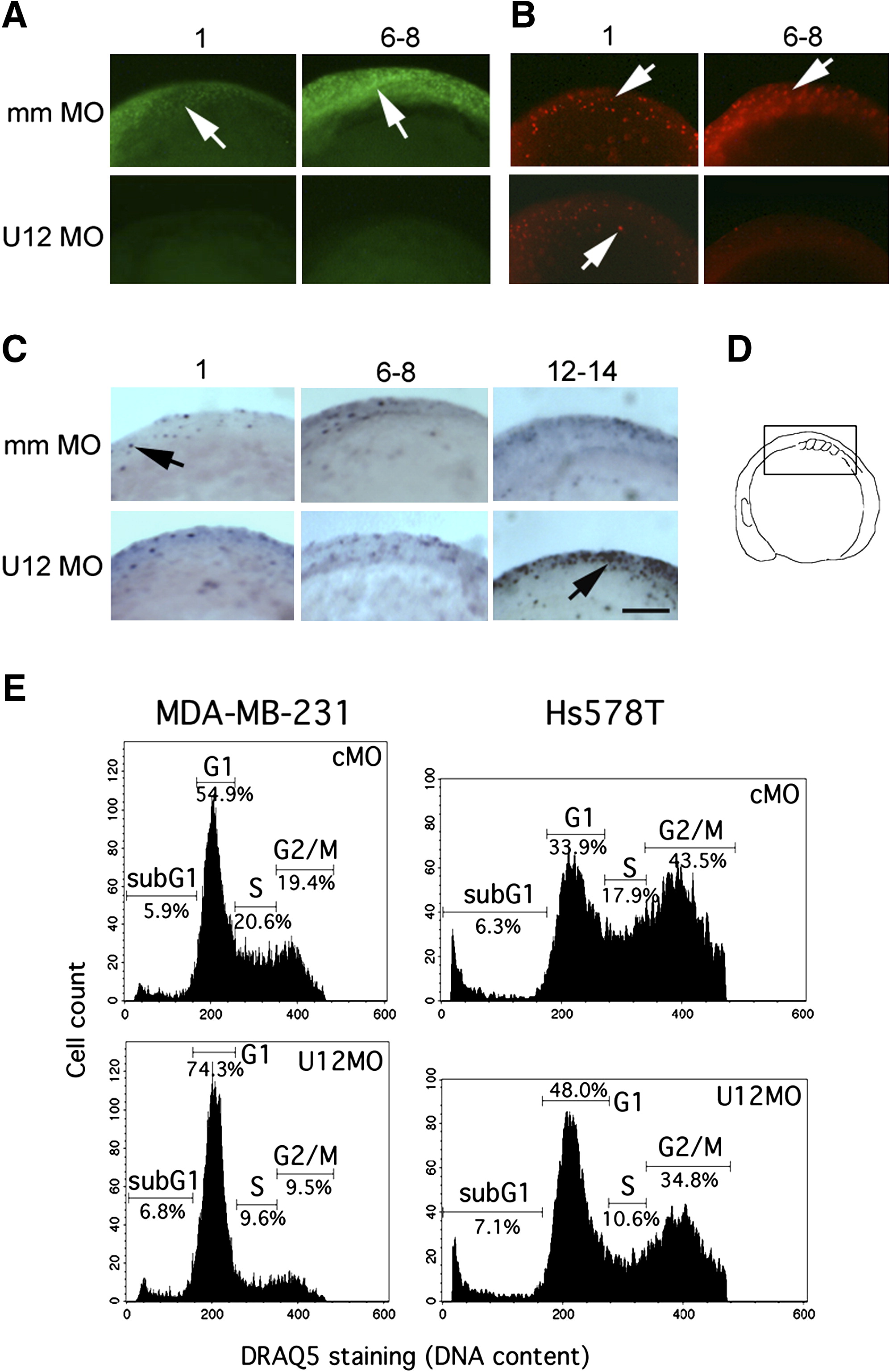
Fig. 5 Interference with Minor Spliceosome Function Blocks Cell-Cycle Progression
(A) BrdU incorporation in mismatch control morpholino-injected embryos (mm MO, 0.1 mM) and U12 morpholino “knockdown” embryos (U12MO, 0.01 mM). BrdU-incorporating cells were visualized by staining with an FITC-conjugated anti-BrdU antibody followed by fluorescence microscopy (arrows).
(B) Analysis of cells in mitosis (M) phase in embryos injected as in (A) by using anti-phospho-histone H3/Cy3 fluorescence staining (arrows).
(C) Detection of apoptosis (arrows) in control- and U12 morpholino-treated embryos by TUNEL staining. Numbers above the panels represent the number of somites in the embryos as indication of their developmental stages. Scale bar indicates 100 μm.
(D) Embryos in all panels are shown in lateral views (anterior to the left) on to the midline at the level of developing somites, as indicated in the schematic drawing.
(E) Morpholino-mediated interference with minor-class splicing inhibits entry into DNA-synthesis (S) phase in human cell lines. Cell-cycle profiles measured by flow cytometry of MDA-MB-231 and Hs578T breast cancer cells after transfection with either a standard control morpholino (cMO) or an antisense-U12 morpholino (U12MO). Cells were ethanol fixed 24 hr after transfection, and cellular DNA was stained with DRAQ5. SubG1 indicates cells with DNA content < 2n.
Reprinted from Cell, 131(4), König, H., Matter, N., Bader, R., Thiele, W., and Müller, F., Splicing Segregation: The Minor Spliceosome Acts outside the Nucleus and Controls Cell Proliferation, 718-729, Copyright (2007) with permission from Elsevier. Full text @ Cell