
|
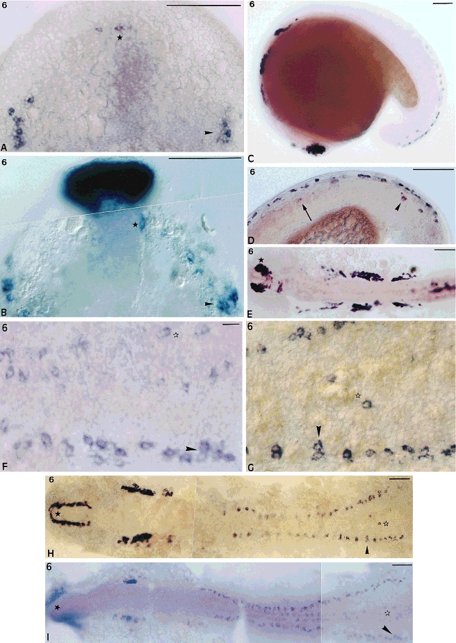
Fig. 6 nrd expression detected by whole-mount in situ hybridization. In Aand B the embryos are positioned such that the anterior end is up, and in all other cases the anterior end is to the left. A: nrd expression in the 10 hours postfertilization (hpf)-embryo, dorsal view of the rostral part of the neural plate. Arrowhead, trigeminal ganglia; star, brain cells. B: islet-1 expression in the rostral part of the neural plate, stage 10 hpf, top view. Arrowhead, trigeminal ganglia; star, brain cells. C: nrd expression in the 17-hpf embryo, side view. D: nrd expression in the caudal region of the 17-hpf zebrafish embryo, side view. Caudal MNs, arrowhead; more rostral MNs, arrow. E: nrd expression in the rostral part of the 22-hpf zebrafish embryo, dorsal view. Asterisk, telencephalon. H: nrd expression in the 12-hpf spread zebrafish embryo, dorsal view. Star, telencephalon; arrowhead, Rohon-Beard cells; open star, motoneurons. G: Arrowhead and open star define same cells as in H. I: islet-1 expression in the 12 hpf spread zebrafish embryo, dorsal view. Star, telencephalon; arrowhead, Rohon-Beard cells; open star, motoneurons. F: Blowup, arrowhead and open star define same cells as in I. Scale bars = 100 μm in A-E,H,I, 10 μm in F,G.