
Fig. 1
- ID
- ZDB-IMAGE-080702-38
- Publication
- Hyatt et al., 1996 - Retinoic acid alters photoreceptor development in vivo
- All Figures
- Figures for Hyatt et al., 1996
|
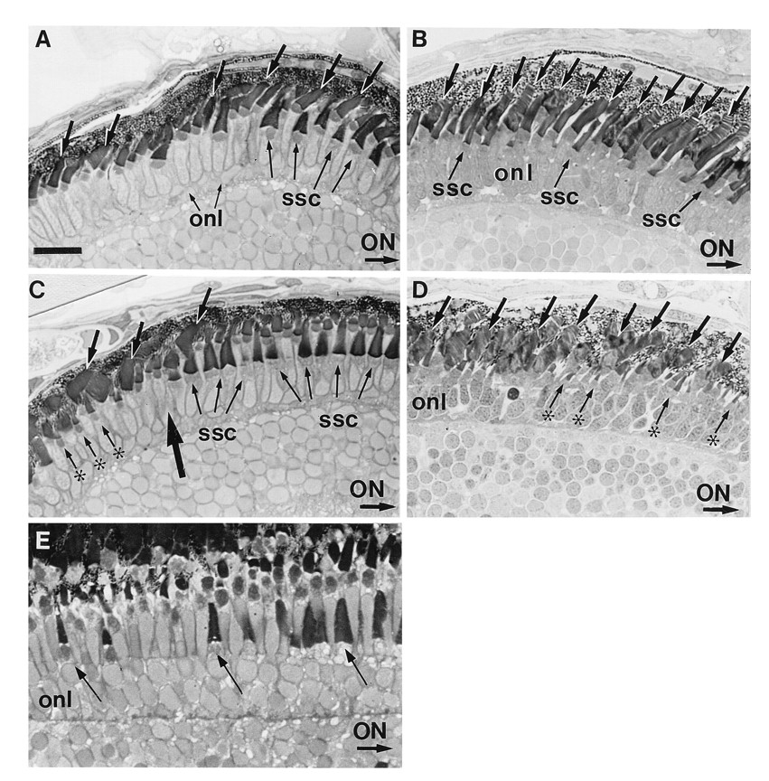
Fig. 1 (A–D) Transverse sections of normal (A and C) and RA-treated (B and D) embryos on day 5 pf. In these sections, the optic nerve (ON) is to the right. (A) In the midperipheral region of the dorsal retina of control embryos, short single cones (ssc) with broad outer segments lie vitreal to rods and longer single cones. The short single cones are more obvious to the right, i.e., more centrally. Rod outer segments average 6 μm in length and are observed across all of this retinal region (arrows). (B) After a 3-day treatment with RA, the number of observable rods (arrows) has increased within the dorsal retina, and the short single cones (ssc) are smaller and less obvious. (C) The ventral retina in control eyes is partitioned into two compartments: a cone-dominated region (right of large arrow) adjacent to the optic nerve and a peripheral region (left of large arrow) dominated by rods (arrows). Mature short single cones (ssc) lie vitreal to a layer of longer single cones in the cone-dominated region. In the rod-dominated region, a population of miniature cones (asterisks) are seen. (D) After RA treatment beginning at day 2 pf, the cone-dominated region within the ventral retina is not observed. Rather large rod outer segments (arrows) are observed extending across the entire ventral region rather than being restricted to the periphery. These rod outer segments appear less organized than those of controls and average ≈10 μm in length, which is ≈2 μm longer than those observed in the ventral region of controls. Small cones (asterisks) resembling the miniature cones located within the ventral periphery in controls extend across the entire ventral region. (E) Transverse section of the peripheral retina from a normal retina at 21 days pf. Arrows identify maturing short single cones (from left to right). onl, Outer nuclear layer. (A–D, bar = 16 μm; E, bar = 9 μm.)