
|
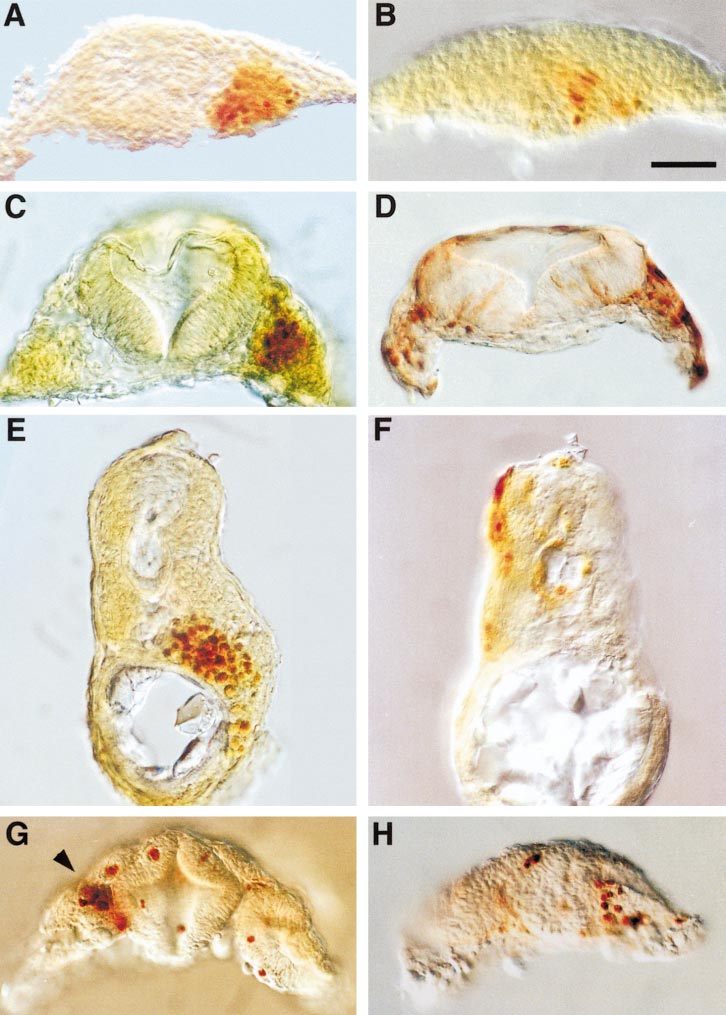
Fig. 4 Appearance of zOtx1-induced cell aggregates. Embryos were injected with myc-zOtx1-wt (A, C, E) or myc-zOtx1-ΔH (B, D, F) into one blastomere of a 16-cell embryo or with myc-zOtx1-wt into one blastomere of a 32-cell embryo (G, H) as in Fig. 5. Embryos were allowed to develop to 14 hpf (A, B) or 24 hpf (C–H), processed for immunohistochemical localization of myc epitope, and hand-cut to obtain transverse sections. myc-Otx1-wt, but not myc-zOtx1-ΔH, induces aggregates in the prospective 14-hpf hindbrain region (A, B), in the mesenchyme flanking rhombomere 3 of a 24-hpf embryo (C, D), and in the midtrunk gut region of a 24-hpf embryo (E, F). In (E) only scattered mesenchymal cells stain positively for myc epitope—the brownish color is due to staining of nonadjacent cells at a deeper layer of the section. In midbrain (G) and forebrain regions (H) of embryos injected with myc-zOtx1 RNA, individual cells or small aggregates of cells are found to express the protein. The section shown in (G) has been obliquely cut through the midbrain so that the most posterior portion of the left neural retina and a more anterior portion of the right neural retina are included. A small aggregate of myc-zOtx1- expressing cells is found in the medial posterior portion of the left neural retina (arrowhead), whereas scattered expressing cells are found in the right neural retina. The section shown in (H) is cut through the telencephalon of another embryo. Scale bar, 50 μm (all at same magnification).
Reprinted from Developmental Biology, 223(2), Bellipanni, G., Murakami, T., Doerre, O.G., Andermann, P., and Weinberg, E.S., Expression of Otx homeodomain proteins induces cell aggregation in developing zebrafish embryos, 339-353, Copyright (2000) with permission from Elsevier. Full text @ Dev. Biol.