
|
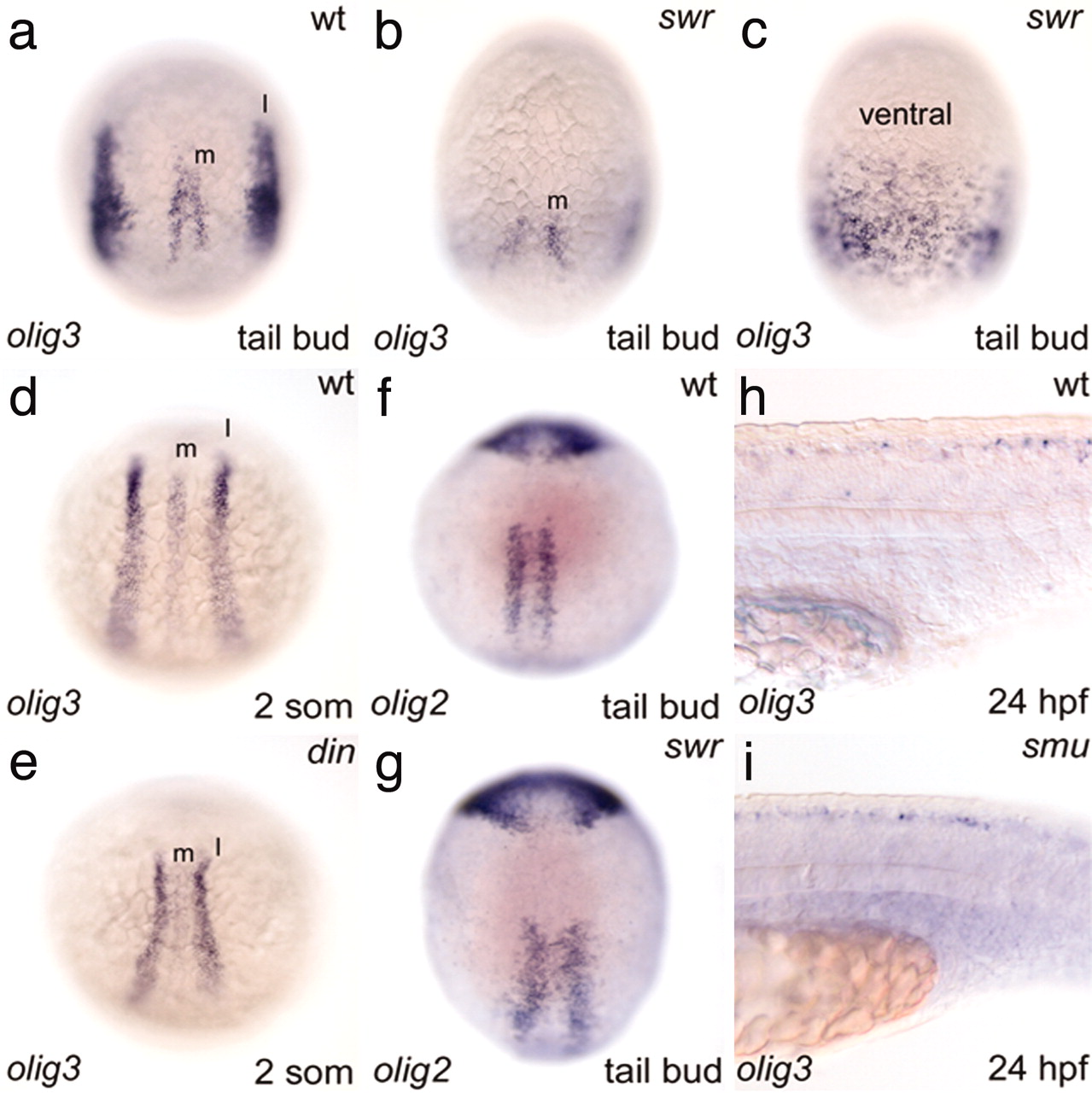
Fig. 6 BMP-dependent restriction of olig3 expression domains. (a) Normal embryo. (b) swr embryo showing a widened space between the two medial domains (m). (c) Ventral view of a swr embryo showing that the lateral olig3 domains are broadened and merge on the ventral side. (d) Normal embryo. (e) din embryo with reduced lateral (l) domain. (f and g) Normal (f) and swr (g) embryos show a similar olig2 pattern. (h and i) Normal expression of olig3 in the dorsal spinal cord of the smu embryo. Embryos in a–g are in dorsal view with anterior to the top, except in c (ventral view). Embryos in h and i are in lateral view, anterior to the left.