
|
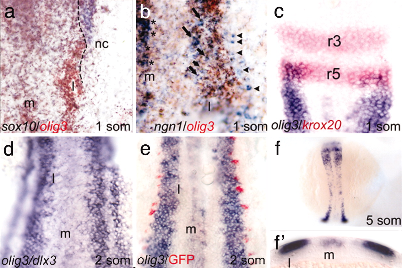
Fig. 1 olig3 expression pattern. (a) High magnification of the right side of the rhomboencephalon region showing that olig3 (brown) is expressed medially to the neural crest marker sox10 (blue); the dashed line represents the boundary of expression of the two markers. (b) High magnification of the right side of the trunk region shows that the lateral (l) olig3 (brown) expression domain comprises the region where ngn1-positive interneurons (arrows) differentiate. RB cells (arrowheads) are in a more lateral position and are outside the olig3 expression domain. (c) The anterior border of the olig3 domain (blue) is delimited by krox20 (red) in rhombomere-5 (r5). (d) The olig3 domain has elongated rostrocaudally, and a two-cell-wide gap separates olig3 and dlx3. (e) ngn1:GFP transgenics showing that RB-cell precursors (red) and olig3-expressing cells are in close contact. (f) At the five-somite stage, the lateral domain has further elongated. (f′) Transverse section at the level of the posterior trunk of the embryo in f. The embryos are in dorsal view, anterior to the top.