
|
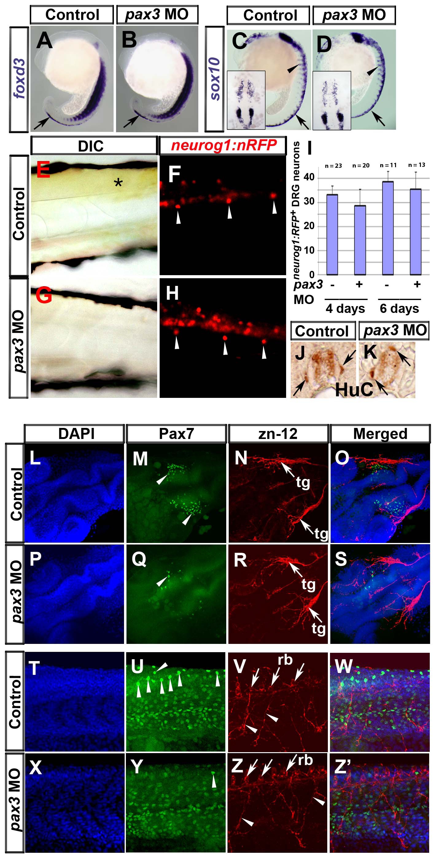
Fig. S2 Pax3 is not required for sensory neural crest lineages. In situ mRNA hybridisation analysis of foxd3 (A,B) and sox10 (C,D), live bright field (E,G) and fluorescent (F,H) analysis of the neurog1:nRFP transgenic fish line and immunodetection of HuC (J,K), Pax7 (M,Q,U,Y) and zn-12 (N,R,V,Z). Lateral view wholemounts are anterior to top and dorsal to right (A-D). Lateral view flatmounts are anterior to left and dorsal to top (E-H, T-Z′). Dorsal view flatmounts are anterior to top (C,D insets) or lower left (L-S). Transverse cryosections are dorsal to top (J,K). A-D. At 18s, foxd3 mRNA is detected in pre-migratory NC (arrows), whereas sox10 mRNA is in both pre-migratory and migrating NC (arrowheads). Expression of foxd3 and sox10 appears unaffected in pax3 morphants (B,D). E-K. In 6 dpf larvae, xanthophore pigmentation is present in the trunk of neurog1:nRFP transgenic fish (E, asterisk), in which DRG neurons (F, arrowheads) and weaker CNS neurons are visible. Injection of pax3 MO ablates xanthophore pigmentation (G) but DRG neurons remain (H, arrowheads). Quantification of DRG neuron number reveals that there is no significant difference between control and pax3 morphants (I). DRG neurons are sometimes mis-positioned dorsally in pax3 morphants (J,K arrows). L-Z′. At 24 hpf, Pax7+ NC cells are clustered in the head of control embryos (M, arrowheads) near to zn-12 immunoreactive trigeminal ganglia (N, arrows, tg). In pax3 morphant, there are fewer Pax7+ NC cells in cranial regions (Q, arrowhead) but the trigeminal ganglia have formed normally (R, arrows). In trunk regions, Pax7+ NC cells are situated in the pre-migratory position (U, arrowheads). Zn-12 staining shows the Rohon-Beard neuronal cell bodies (V, arrows, rb) and processes (V, arrowheads). Pax3 morphants lack Pax7+ pre-migratory NC cells (Y), whereas Rohon-Beard neuronal cell bodies and processes (Z) and dermomyotomal Pax7 accumulation (Y) appear normal.
Reprinted from Developmental Biology, 317(2), Minchin, J.E., and Hughes, S.M., Sequential actions of Pax3 and Pax7 drive xanthophore development in zebrafish neural crest, 508-522, Copyright (2008) with permission from Elsevier. Full text @ Dev. Biol.