
|
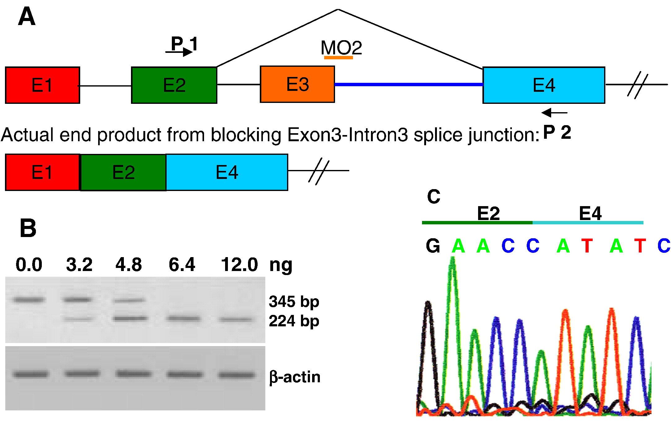
Fig. 3 Validation of ndrg4-MO2 (knockdown by splice-blocking). (A) Schematic of the strategy to target the exon3–intron3 boundary with ndrg4-MO2. Arrows represent primers used in the RT-PCR to test the efficiency of ndrg4 splice-blocking MO2 and the orange bar represents the ndrg4-MO2. (B) The RT-PCR was performed on wild-type embryos and embryos injected with 3.2–12.0 ng of the ndrg4-MO2. The ndrg4-MO2 efficiently blocks ndrg4 mRNA splicing by exon3 deletion in a dose-dependent manner. Bands of 345 bp and 224 bp represent wildtype and exon3-deleted ndrg4 mRNA, respectively. (C) Sequencing of the 224 bp band reveals direct connection of exon 2 and exon 4.
Reprinted from Developmental Biology, 317(2), Qu, X., Jia, H., Garrity, D.M., Tompkins, K., Batts, L., Appel, B., Zhong, T.P., and Baldwin, H.S., ndrg4 is required for normal myocyte proliferation during early cardiac development in zebrafish, 486-496, Copyright (2008) with permission from Elsevier. Full text @ Dev. Biol.