
Image
|
Figure Caption
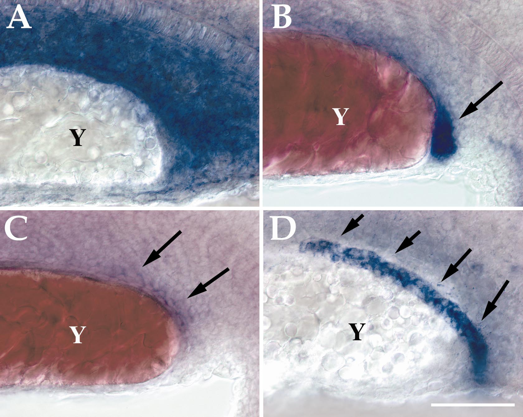
Fig. 3 Pronephric duct and mesenchyme mRNA expression of zebrafish GDNF, GFRα1, and RET at 20 hpf. (A–D) Lateral view of the ventral half of the trunk of 18 hpf embryos from somite 10–18 that have been hybridized with riboprobes for zebrafish GDNF (A), GFRα1a (B), GFRα1b (C), and RET (D) messages. Y, yolk sac extension. Arrow in (B) indicates the posterior part of the pronephric duct. Arrows in (C) indicate weak GFRα1b expression in the pronephric duct. Arrows in (D) indicate pronephric duct. Rostral is to the left. Scale bar: 50 μm.
Figure Data
Acknowledgments
This image is the copyrighted work of the attributed author or publisher, and
ZFIN has permission only to display this image to its users.
Additional permissions should be obtained from the applicable author or publisher of the image.
Reprinted from Developmental Biology, 231(2), Shepherd, I.T., Beattie, C.E., and Raible, D.W., Functional analysis of zebrafish GDNF, 420-435, Copyright (2001) with permission from Elsevier. Full text @ Dev. Biol.