
|
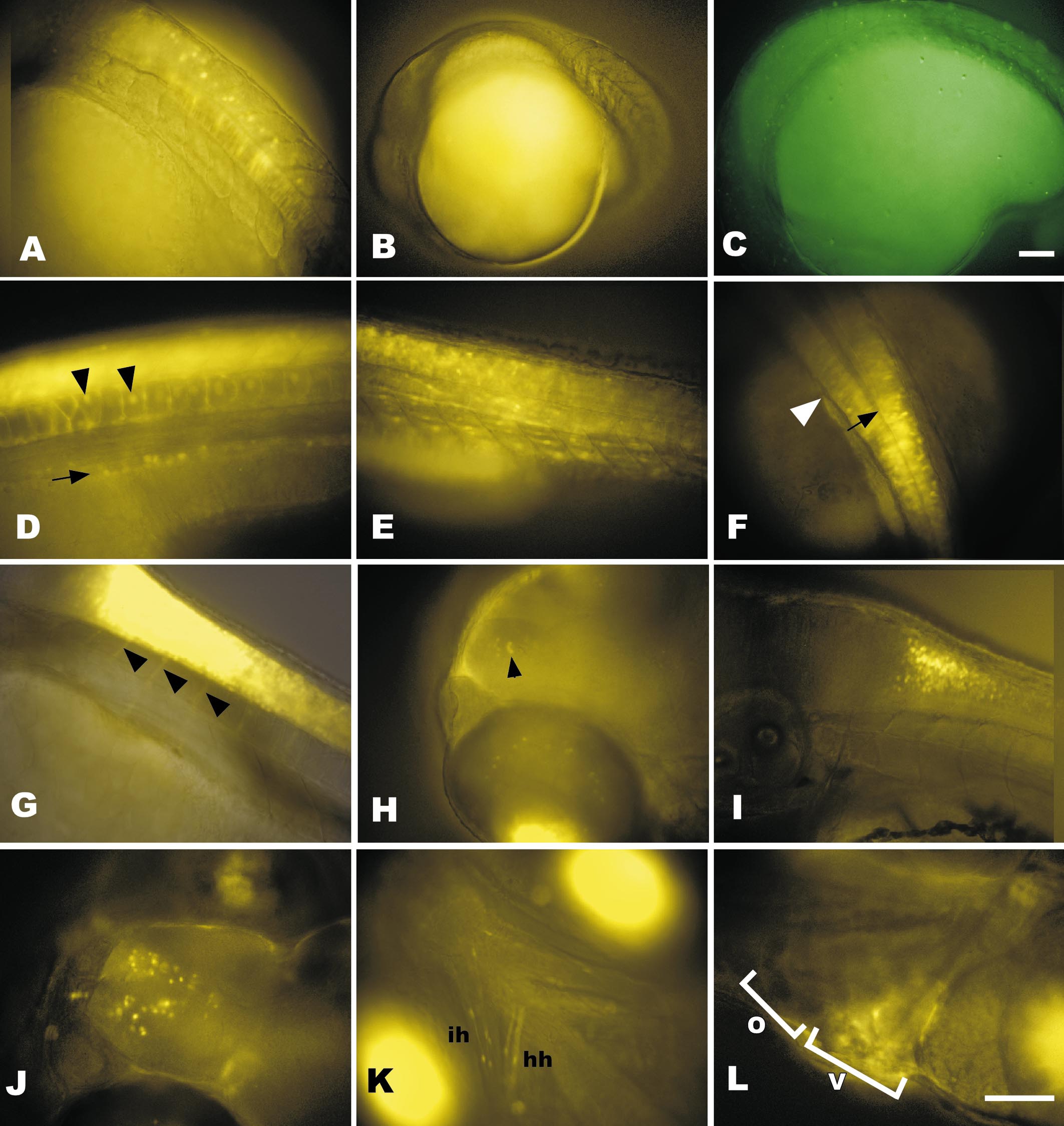
Fig. 4 RGY expression patterns are more complex than RTG expression. (A) Notochord and neural tube expression in RGY at 18 hpf is in a subset of cells in each tissue. (B and C) Fluorescent images of nontransgenic embryos at 18 hpf to demonstrate typical autofluorescence when using long exposure times required to capture early transgene expression shown in Figs. 2A and 2B with yellow (B) or green (C) filter sets. Exposure times were 5 s. (D–F) Transgene expression in 30 hpf RGY embryos in the pronephric ducts (arrow) and vacuolated notochord (arrowheads) at the level of the junction between yolk and yolk extension (D). Expression also occurs in the somites (E). In the neural tube (shown in dorsal view in F) expression extends rostrally into the caudal hindbrain beyond the posterior tip of the hindbrain ventricle (black arrow) and first myotome (white arrowhead). (G) At 48 hpf neural tube expression extends ventrally in axon-like projections to the somites (arrowheads). (H) Dorsolateral view of rostral end of 72 hpf RGY larvae showing an arch of expression in cells of the inner nuclear layer of the dorsal retina and a small patch of forebrain expression (arrowhead). Bright fluorescence at the bottom of the image is the ventral retina. (I) Lateral view of 72-hpf hindbrain/spinal cord junction imaged as a confocal slice shows higher density of dorsal cells expressing the transgene than ventral. (J) Dorsal view of expression in the forebrain at 5 days postfertilization. (K) Ventral view of transgene expression in cranial skeletal muscles, hyohyoideus (hh) and interhyodoideus (ih). Bright bilateral fluorescence is expression in the ventral retina. (L) Lateral view of 5-day heart showing blurred expression in the beating ventricle (V) that is excluded from the outflow tract (O). A–E, G–I, and L are lateral views with rostral left and dorsal up. F is a dorsal view with rostral up. J and K are dorsal and ventral views, respectively, with rostral left. Bars, 100 μm. Scale for A and D–K is the same as in L. Scale for B is same as in C.
Reprinted from Developmental Biology, 229(1), Perz-Edwards, A., Hardison, N.L., and Linney, E., Retinoic acid-mediated gene expression in transgenic reporter zebrafish, 89-101, Copyright (2001) with permission from Elsevier. Full text @ Dev. Biol.