
|
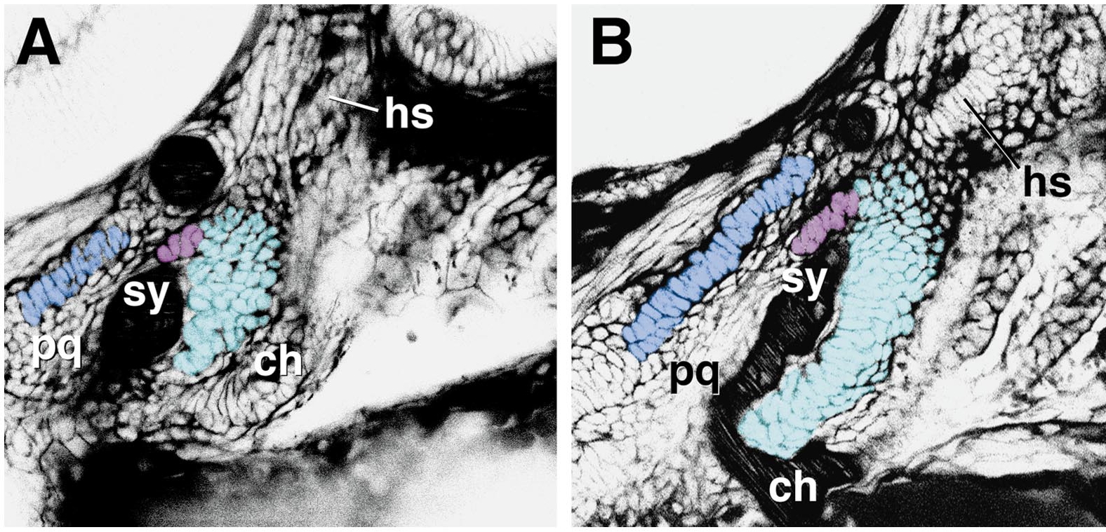
Fig. 10 Morphogenesis (chondrogenesis) in the anterior pharyngeal wall, as revealed by BODIPY ceramide labeling in live WT preparations (left side view). (A) 52 h, (B) 58 h (unpublished confocal micrographs, M. Jones and C.B.K.). Negative images are shown, allowing the various regions of developing cartilages to be pseudocolored for clarity of presentation. The mature cartilages are shown in the same orientation in Fig. 1D. Here the two distinctive regions of the dorsal hyoid cartilage (the hyosymplectic, Fig. 1) are indicated separately, the strut-like symplectic region (sy) and the plate-like hyomandibular region (hm). The more dorsal part (hyomandibular region; for more detail see Kimmel et al., 1998) is left uncolored. Both of these cartilage-forming regions, as well as the ventral cartilage in this segment, the ceratohyal (ch), are developing out of a common primordium, the hyoid condensation. In contrast, the palatoquadrate (pq) and Meckel’s cartilage (only partly included here) can be seen to be developing out of another shared primordium, the mandibular condensation, as described previously from analysis of fixed material (Schilling and Kimmel, 1997; see also Fig. 8).
Reprinted from Developmental Biology, 233(2), Kimmel, C.B., Miller, C.T., and Moens, C.B., Specification and morphogenesis of the zebrafish larval head skeleton, 239-257, Copyright (2001) with permission from Elsevier. Full text @ Dev. Biol.