
|
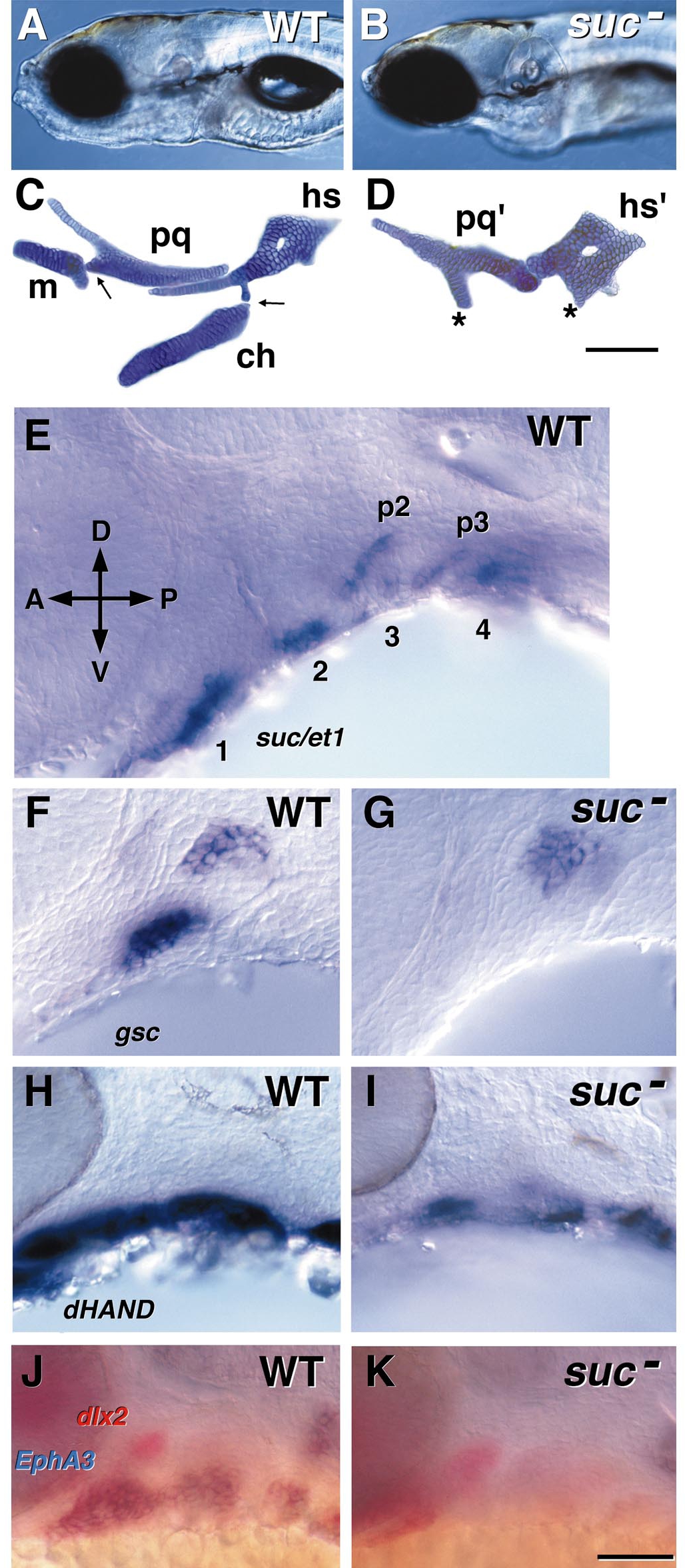
Fig. 7 Phenotype and gene expression in WT and suc mutants (from Miller et al., 2000). WT are to the left and suc mutants to the right. (A, B) The craniofacial appearance in left side view of WT and a suc mutant at day 4. (C, D) The cartilage phenotype at day 4. Alcian blue-labeled, flat-mounted elements of the mandibular and hyoid arches (pharyngeal segments 1 and 2). The abbreviations are as in Fig. 1. The asterisks in D show regions of ventral cartilage that may correspond to m and ch in the first and second segments, respectively. Scale bar, 100 μm. (E) Whole mount RNA in situ preparation (36 h, left side view) showing segmental expression of suc/et-1 in the ventralpharyngeal arches. The first four segments are indicated (1– 4), and labeling is also present in endodermal pouches 2 and 3 (p2, p3). (F–K) Targets of suc signaling, as revealed by comparing the RNA expression patterns in WT and suc mutants. Left side views of whole mounted embryos. (F, G) goosecoid (gsc), 30 h. (H, I) dHAND, 28 h. (J, K) dlx2 (in red) and EphA3 (in blue), 32 h. Scale bar (K), 50 μm.
Reprinted from Developmental Biology, 233(2), Kimmel, C.B., Miller, C.T., and Moens, C.B., Specification and morphogenesis of the zebrafish larval head skeleton, 239-257, Copyright (2001) with permission from Elsevier. Full text @ Dev. Biol.