
|
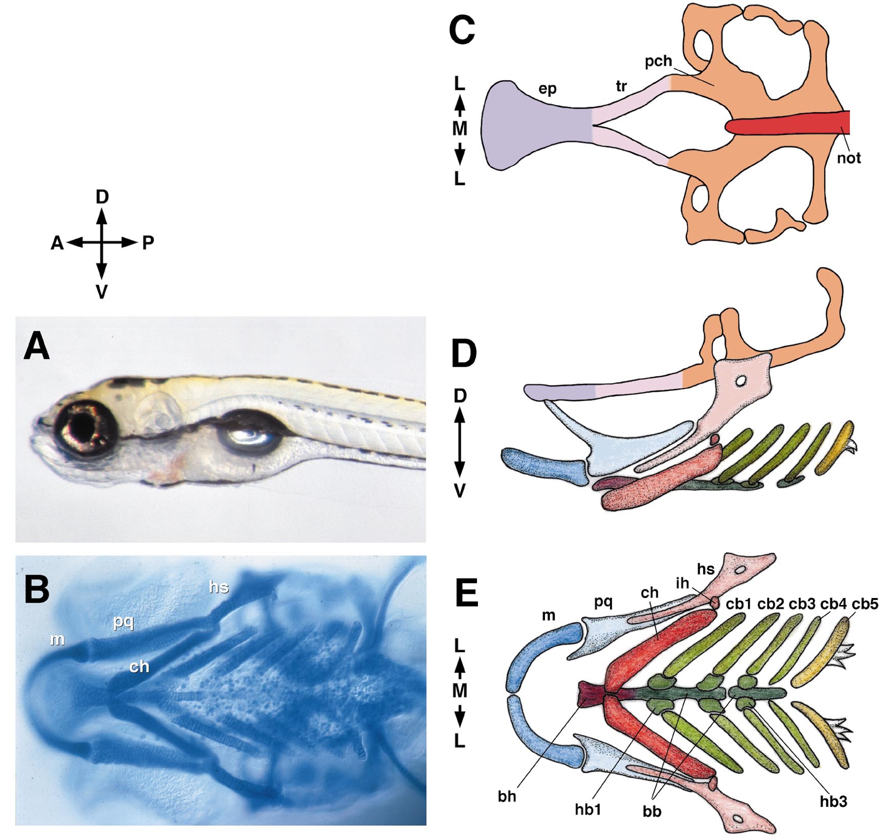
Fig. 1 The young larval zebrafish (A, 5 days postfertilization, left side view) and the layout of its cartilaginous head skeleton. For a further description see Schilling and Kimmel (1997) and Cubbage and Mabee (1996). Anterior is to left in this and other figures in the paper except where indicated. B shows a ventral view of an Alcian blue-labeled, whole-mounted preparation. Cartilages are indicated in the first or mandibular arch (pq, m) and in the second or hyoid arch (hs, ch). C–E show drawings of such preparations. (C) The neurocranial (or basicranial) cartilages and notochord from the dorsal aspect. The eyes fit into the shallow grooves along the sides of the ethmoid plate and trabeculae. The otic vesicles fit into the prominent cavities to either side of the notochord and parachordal cartilages . The brain’s posterior pituitary fits into the prominent midline cavity ahead of the notochord, the hypophysial fenestra. (D). The neurocranium (diagrammatically elevated dorsalward for the sake of clarity) and the pharyngeal skeleton in side view. (E). The pharyngeal skeleton in ventral view. Abbreviations used: A, anterior; bb, basibranchial; bh, basihyal; cb, ceratobranchial; ch, ceratohyal; D, dorsal; ep, ethmoid plate; hb, hypobranchial; hs, hyosymplectic; ih, interhyal; L, lateral; M, medial; m, Meckel’s; not, notochord; P, posterior; pch, parachordal; pq, palatoquadrate; tr, trabecula; V, ventral.
Reprinted from Developmental Biology, 233(2), Kimmel, C.B., Miller, C.T., and Moens, C.B., Specification and morphogenesis of the zebrafish larval head skeleton, 239-257, Copyright (2001) with permission from Elsevier. Full text @ Dev. Biol.