
Image
|
Figure Caption
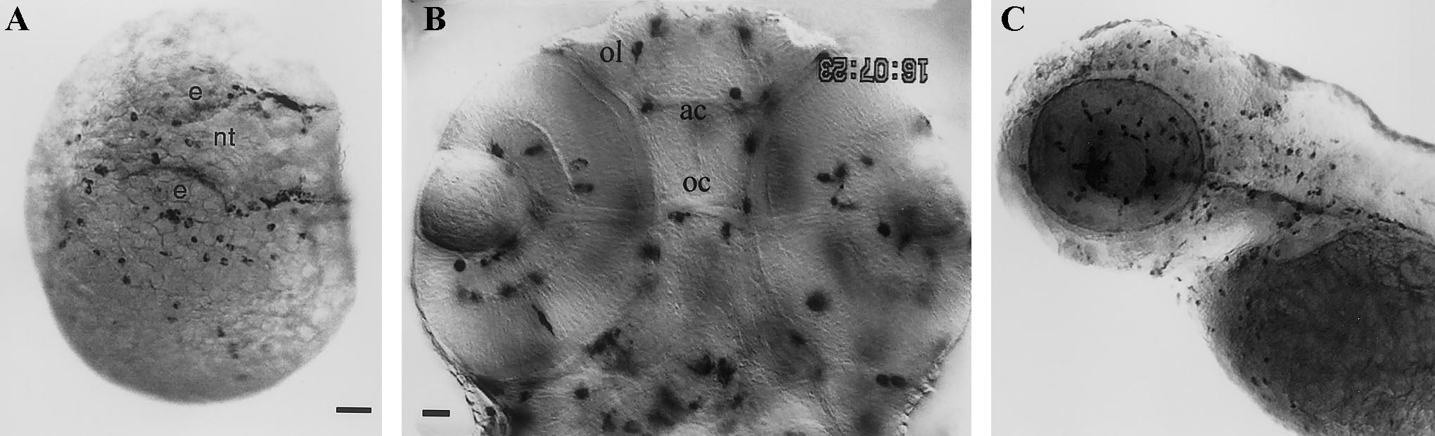
Fig. 8 Expression of the fms gene in the early macrophage lineage. (A) 25-somite (21.5 hpf), dorso-lateral view; beside pre-/young macrophages spread on the yolkball, fms-positive xanthophore precursors outline the lateral borders of the neural tube (nt); (e) eye. (B) 48 hpf, head, deep dorsal view. Out-of-focus fms-positive macrophages produce the multiple shadows. (ol) olfactory organ; (ac) anterior commissure; (oc) optic chiasm. (C) 72 hpf, lateral view. Bars: (A, C) 50 μm; (B) 20 μm.
Acknowledgments
This image is the copyrighted work of the attributed author or publisher, and
ZFIN has permission only to display this image to its users.
Additional permissions should be obtained from the applicable author or publisher of the image.
Reprinted from Developmental Biology, 238(2), Herbomel, P., Thisse, B., and Thisse, C., Zebrafish early macrophages colonize cephalic mesenchyme and developing brain, retina, and epidermis through a M-CSF receptor-dependent invasive process, 274-288, Copyright (2001) with permission from Elsevier. Full text @ Dev. Biol.