
|
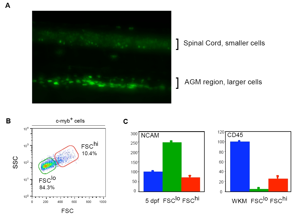
Fig. S2 HSCs and neurons can be segregated by light scatter characteristics from cmyb:eGFP embryos. (A) Lateral view of a cmyb:eGFP embryo at 42 hpf. The spinal cord shows small round GFP+ cells, whereas larger and brighter cells are observed in the AGM region (dorsal side upwards, anterior towards the left). (B) cmyb:EGFP+ cells were analyzed by forward (FSC) and side (SSC) scatter characteristics. Cells were sorted by size (red gate, larger; green gate, smaller). (C) QPCR analysis shows that FSClo cells are highly enriched for NCAM-expressing cells, whereas FSChi cells are enriched for CD45-expressing cells.