
Fig. 7
|
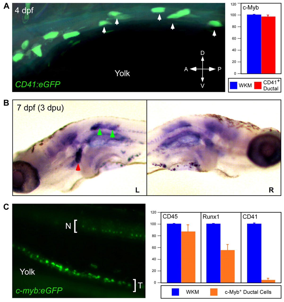
Fig. 7 Characterization of hematopoietic precursors on the pronephric tubules. (A) FITC was uncaged in 5 CD41:eGFP+ cells (arrows) at 4 dpf along the left pronephric tubule. CD41:eGFP+ cells express cmyb (right panel). (B) Animals were fixed 3 days post-uncaging and analyzed for uncaged FITC. Ductal cells migrated from targeted region on the left pronephric tubule (green arrowheads, left panel) to the left anterior pronephros (red arrowhead). Contralateral anterior pronephri were not colonized (right panel). (C) cmyb:eGFP+ tubular cells (T) were purified away from GFP+ neural cells (N) from 75 hpf dissected trunks by flow cytometry and analyzed for hematopoietic gene expression (right panel).