
|
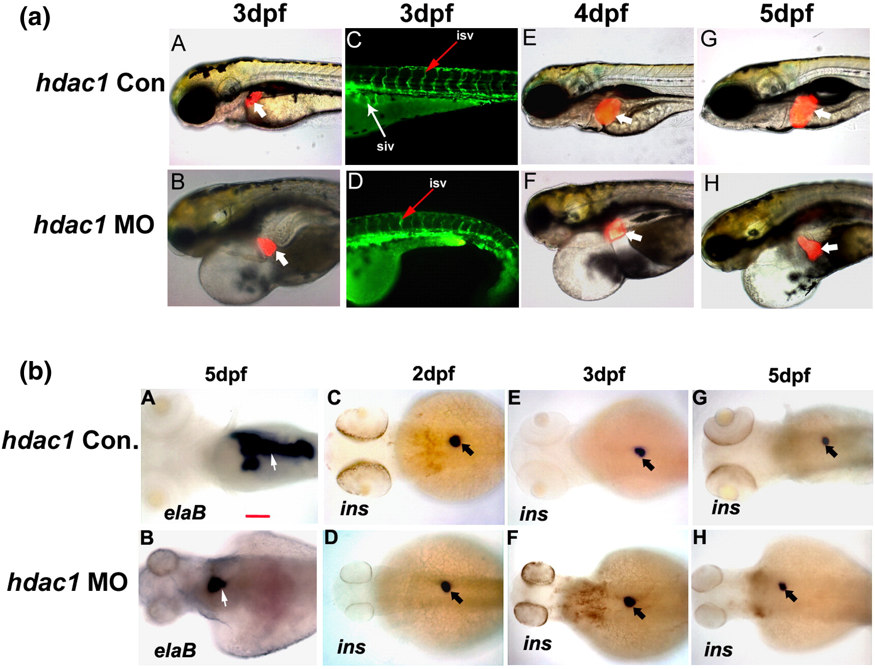
Fig. 6 Role of hdac1 in zebrafish liver, pancreas and blood vessels development. (a) Liver development in hdac1 morphants (6 ng/embryo MO) was analyzed in Tg(lfabp:RFP; elaA:EGFP) from 3 dpf to 5 dpf. Control embryos were injected with the same amount of hdac1 5 bp mismatch MO. Liver (thick white arrow) formed relatively normal in hdac1 morphants at 3 dpf (A, B). However, its subsequent growth was reduced compared to control embryos (E–H). Angiogenesis was analyzed in Tg(fli-1:EGFP) at 3 dpf. SIVs were absent (thin white arrow, D) while ISV defects were mild and rare (thin red arrow, C and D). hdac1 morphants exhibited global embryonic defects with severe cardiac edema, small head and eyes as well as absence of craniofacial cartilage structures (B–H). Scale bar is 100 μm. (b) Zebrafish pancreas development in hdac1 morphants was analyzed by elastase B (A, B) and insulin (C–H) expression through WISH in hdac1 morphants with 5 bp mismatch MO injected embryos as control (Con.). Exocrine pancreas (white arrow) was significantly reduced at 5 dpf (A, B) but endocrine pancreas formation (black arrow) was not affected (C–H). All images are dorsal views, anterior to the left. Scale bar is 100 μm.
Reprinted from Developmental Biology, 317(1), Farooq, M., Sulochana, K.N., Pan, X., To, J., Sheng, D., Gong, Z., and Ge, R., Histone deacetylase 3 (hdac3) is specifically required for liver development in zebrafish, 336-353, Copyright (2008) with permission from Elsevier. Full text @ Dev. Biol.