
Fig. 6
- ID
- ZDB-IMAGE-080417-33
- Publication
- Kotani et al., 2008 - misty somites, a maternal effect gene identified by transposon-mediated insertional mutagenesis in zebrafish that is essential for the somite boundary maintenance
- All Figures
- Figures for Kotani et al., 2008
|
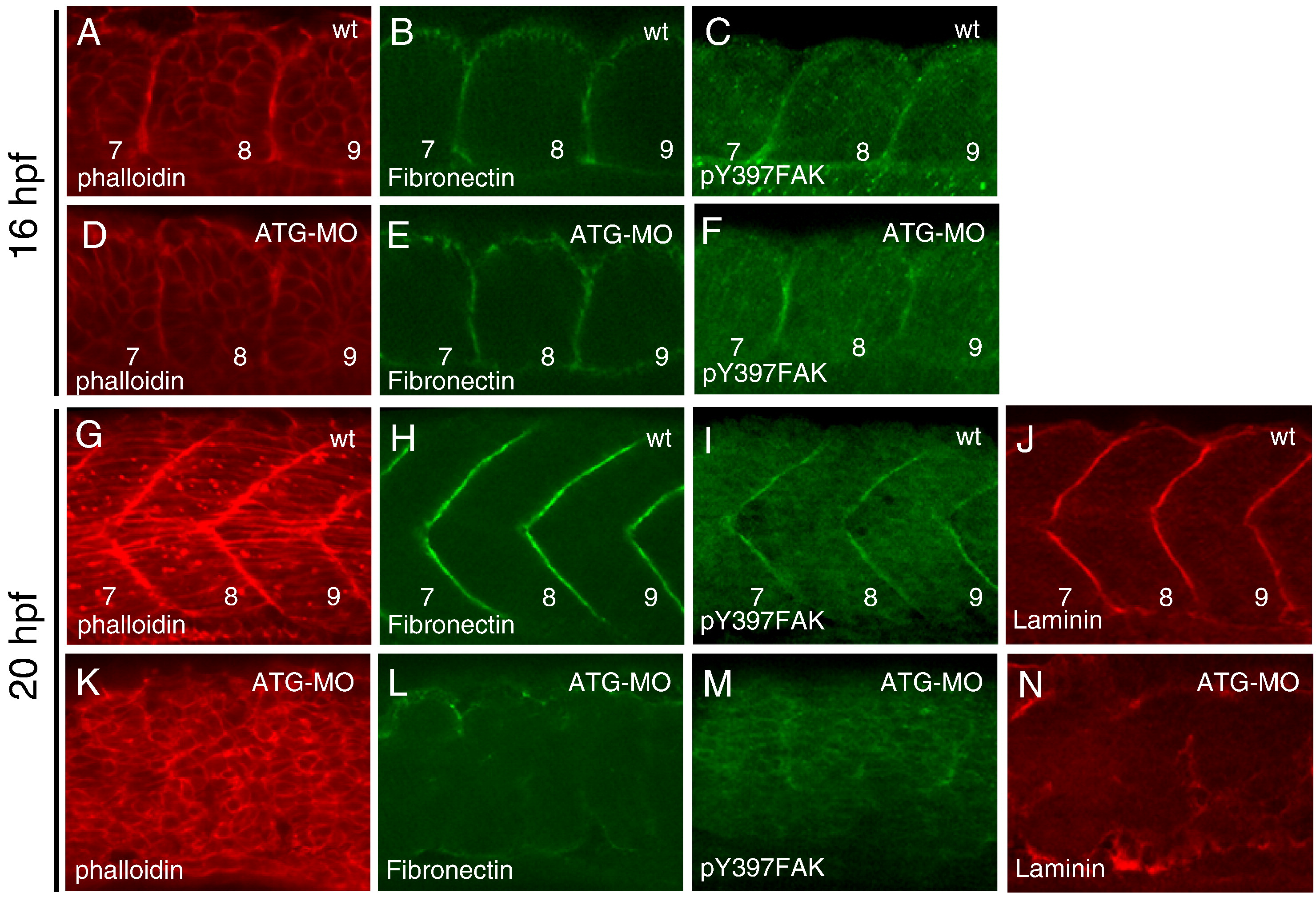
Fig. 6 Disappearance of the somite boundaries in the mys-deficient embryo. Confocal images of wild type (A–C, G–J) and ATG-MO injected embryos (D–F, K–N). (A–F) Dorsal views of the 7th to 9th somite at 16 hpf (the 14-somite stage). (G–N) Lateral views of fast muscle cells of the 7th to 9th somite at 20 hpf (the 21-somite stage). Embryos were stained with phalloidin (A, D, G, K), anti-Fibronectin antibody (B, E, H, L), anti-phosphorylated FAK antibody (C, F, I, M) and anti-Laminin antibody (J, N). Numbers indicate the somite numbers. In the ATG-MO injected embryos, the somite boundaries disappeared at 20 hpf.
Reprinted from Developmental Biology, 316(2), Kotani, T., and Kawakami, K., misty somites, a maternal effect gene identified by transposon-mediated insertional mutagenesis in zebrafish that is essential for the somite boundary maintenance, 383-396, Copyright (2008) with permission from Elsevier. Full text @ Dev. Biol.