
Image
|
Figure Caption
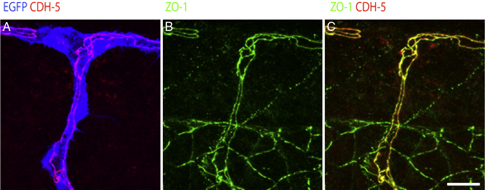
Fig. 3 CDH5 colocalizes with ZO-1 in endothelial cells. (A–C) Confocal projections of 36 hpf fli1:EGFP (blue) transgenic embryos labelled with anti CDH5 (red) and anti ZO-1 (green) antibodies. (A) CDH5 and the GFP show that CDH5 labels exclusively junctions of endothelial cells. (B) ZO-1 labels endothelial cells as well as cells in the notochord and the neural tube. (C) CDH5 and ZO-1 show co-localization (yellow) of the two junctional proteins in endothelial cells. Scalebars: 20 μm.
Figure Data
Acknowledgments
This image is the copyrighted work of the attributed author or publisher, and
ZFIN has permission only to display this image to its users.
Additional permissions should be obtained from the applicable author or publisher of the image.
Reprinted from Developmental Biology, 316(2), Blum, Y., Belting, H.G., Ellertsdottir, E., Herwig, L., Lüders, F., and Affolter, M., Complex cell rearrangements during intersegmental vessel sprouting and vessel fusion in the zebrafish embryo, 312-322, Copyright (2008) with permission from Elsevier. Full text @ Dev. Biol.